Sensory Modality Is Determined by the Stimulus Energy Since ancient times five major sensory modalities have been recognized: vision, hearing, touch, taste, and smell. In addition to these classical senses we also consider the somatic senses of pain, temperature, itch, and proprioception (posture and the movement of parts of the body) and the vestibular sense of balance (the position of the body in the gravitational field). An early insight into the neuronal basis of sensation came in 1826, when Johannes Müller advanced his “laws of specific sense energies.” Müller proposed that modality is a property of the sensory nerve fiber. Each nerve fiber is activated primarily by a certain type of stimulus and each makes specific connections to structures in the central nervous system whose activity gives rise to specific sensations. Thus Müller's laws of specific sense energies identified the most important mechanism for neural coding of stimulus modality. The Sensory Neurons for Hearing, Taste, and Smell Are Spatially Organized According to Sensitivity For hearing and the chemical senses (taste and smell), the receptors are spatially distributed following the energy spectrum for these modalities. For example, auditory receptors are arranged according to the sound frequencies to which they respond. Receptors at a specific location vibrate most strongly when stimulated by a particular range of sounds, with high frequencies located at the base of the cochlea and low frequencies at the apex. Thus the organization of the inner ear's receptor sheet represents the spectrum of sound, not the location of the sounds in space. For taste and smell, receptors that have particular chemical sensitivities are located in different parts of the receptive surface of the tongue and inside the nose. For example, specific regions of the tongue contain receptors sensitive to salts, sugars, acids, bases, or proteins. Different foods will excite specific combinations of these receptors to evoke their characteristic tastes. The spatial distribution of activity in the chemoreceptor population allows the brain to differentiate salty from sweet or bitter tastes.
Figure 21-1 The sensory systems encode four elementary attributes of stimuli modality, location, intensity, and timing—which are manifested in sensation. The four attributes of sensation are illustrated in this figure for the somatosensory modality of touch. A. In the human hand the submodalities of touch are sensed by four types of mechanoreceptors. Specific tactile sensations occur when distinct types of receptors are activated. Firing of all four receptors produces the sensation of contact with an object. Selective activation of Merkel cells and Ruffini endings produces sensations of steady pressure on the skin above the receptor. When the same patterns of firing occur only in Meissner's and Pacinian corpuscles, the tingling sensation of vibration is perceived. B. Location and other spatial properties of a stimulus are encoded by the spatial distribution of the population of activated receptors. Each receptor fires action potentials only when the skin close to its sensory terminals is touched, ie, when a stimulus impinges on the receptor's receptive field (see Figure 21-5). The receptive fields of mechanoreceptors—shown as red areas on the finger tip—differ in size and response to touch. Merkel cells and Meissner's corpuscles provide the most precise localization of touch, as they have the smallest receptive fields and are also more sensitive to pressure applied by a small probe. C. The intensity of stimulation is signaled by the firing rates of individual receptors, and the duration of stimulation is signaled by the time course of firing. The spike trains below each finger indicate the action potentials evoked by pressure from a small probe at the center of the receptive field. Two of these receptors (Meissner's and Pacinian corpuscles) adapt rapidly to constant stimulation, while the other two adapt slowly (see Figure 21-8). Sensory Systems Mediate Four Attributes of a Stimulus That Can Be Correlated Quantitatively With a Sensation The modern study of sensation began in the nineteenth century with the pioneering work of Weber and Fechner in sensory psychophysics. They discovered that despite the diversity of sensations we experience, all sensory systems convey four basic types of information when stimulated—modality, location, intensity, and timing. Together, these four elementary attributes of a stimulus yield sensation. The fact that all sensory systems convey the same type of information may be one reason why they have such similar organization. The four fundamental attributes of sensory experience are encoded within the nervous system by specialized subgroups of neurons. Modality defines a general class of stimulus, determined by the type of energy transmitted by the stimulus and the receptors specialized to sense that energy (Figure 21-1). Receptors, together with their central pathways and target areas in the brain, comprise a sensory system, and activity within a system gives rise to specific types of sensations such as touch, taste, vision, or hearing. The location of the stimulus is represented by the set of sensory receptors within the sensory system that are active. Receptors are distributed topographically in a sense organ so that their activity signals not only the modality of the stimulus but also its position in space and its size. As a stimulus activates many receptors simultaneously, the distribution of the active population provides important information to the brain about sensation The intensity of the stimulus is signaled by the response amplitude of each receptor, which reflects the total amount of stimulus energy delivered to the receptor. The timing of stimulation is defined by when the response in the receptor starts and stops and is determined by how quickly the energy is received or lost by the receptor. Therefore, both the intensity and time course of stimulation are represented by the firing patterns of active sensory neurons.
Here is Müller's statement of the law, from Handbuch der Physiologie des Menschen für Vorlesungen , 2nd Ed., translated by Edwin Clarke and Charles Donald O'Malley: The same cause, such as electricity, can simultaneously affect all sensory organs, since they are all sensitive to it; and yet, every sensory nerve reacts to it differently; one nerve perceives it as light, another hears its sound, another one smells it; another tastes the electricity, and another one feels it as pain and shock. One nerve perceives a luminous picture through mechanical irritation, another one hears it as buzzing, another one senses it as pain. . . He who feels compelled to consider the consequences of these facts cannot but realize that the specific sensibility of nerves for certain impressions is not enough, since all nerves are sensitive to the same cause but react to the same cause in different ways. . . (S)ensation is not the conduction of a quality or state of external bodies to consciousness, but the conduction of a quality or state of our nerves to consciousness, excited by an external cause. [edit] Clarification As the above quotation shows, Müller's law seems to differ from the modern statement of the law in one key way. Müller attributed the quality of an experience to some specific quality of the energy in the nerves. For example, the visual experience from light shining into the eye, or from a poke in the eye, arises from some special quality of the energy carried by optic nerve, and the auditory experience from sound coming into the ear, or from electrical stimulation of the cochlea, arises from some different, special quality of the energy carried by the auditory nerve. In 1912, Lord Edgar Douglas Adrian showed that all neurons carry the same energy, electrical energy in the form of action potentials. That means that the quality of an experience depends on the part of the brain to which nerves deliver their action potentials (e.g., light from nerves arriving at the visual cortex and sound from nerves arriving at the auditory cortex). In 1945, Roger Sperry showed that it is the location in the brain to which nerves attach that determines experience. He studied amphibians whose optic nerves cross completely, so that the left eye connects to the right side of the brain and the right eye connects to the left side of the brain. He was able to cut the optic nerves and cause them to regrow on the opposite side of the brain so that the left eye now connected to the left side of the brain and the right eye connected to the right side of the brain. He then showed that these animals made the opposite movements from the ones they would have made before the operation. For example, before the operation, the animal would move to the left to get away from a large object approaching from the right. After the operation, the animal would move to the right in response to the same large object approaching from the right. Sperry showed similar results in other animals including mammals (rats), this work contributing to his Nobel Prize in 1981.
Modality Is Encoded by a Labeled Line Code In each sensory system the initial contact with the external world occurs through specialized neural structures called sensory receptors. The sensory receptor is the first cell in each sensory pathway and transforms stimulus energy into electrical energy, thus establishing a common signaling mechanism in all sensory systems. The electrical signal produced by the receptor is termed the receptor potential. The amplitude and duration of the receptor potential are related to the intensity and time course of stimulation of the particular receptor. The process by which specific stimulus energy is converted into an electrical signal is called stimulus transduction. Receptors are morphologically specialized to transduce specific forms of energy. Each receptor has a specialized anatomical region where stimulus transduction occurs. Most sensory receptors are optimally selective for a single stimulus energy, a property termed receptor specificity. The unique stimulus that activates a specific receptor at a low energy level was called an adequate stimulus by Charles Sherrington. The specificity of response in receptors underlies the labeled line code , the most important coding mechanism for stimulus modality. The fact that the receptor is selective for a particular type of stimulus energy means that the axon of the receptor functions as a modality-specific line of communication; activity in the axon necessarily conveys information about a particular type of stimulus. Excitation of a particular sensory neuron, whether naturally or artificially by direct electrical stimulation, elicits the same sensation. For example, electrical stimulation of the auditory nerve can be used to signal tones of different frequencies in patients with deafness caused by damage to receptors in the inner ear. Each class of sensory receptors makes connections with distinctive structures in the central nervous system, at least in the early stages of information processing. Thus, sight or touch is experienced because a particular central nervous structure is activated. Modality is therefore represented by the ensemble of neurons connected to a specific class of receptors. Such ensembles of neurons are referred to as sensory systems and comprise the somatosensory system, visual system, auditory system, vestibular system, olfactory system, and gustatory system. Transduction of Sensory Stimuli into Nerve Impulses Mechanisms of Receptor Potentials. Different receptors can be excited in one of several ways to cause receptor potentials: by mechanical deformation of the receptor, which stretches the receptor membrane and opens ion channels; by application of a chemical to the membrane, which also opens ion channels; by change of the temperature of the membrane, which alters the permeability of the membrane; or by the effects of electromagnetic radiation, such as light on a retinal visual receptor, which either directly or indirectly changes the receptor membrane characteristics and allows ions to flow through membrane channels. It will be recognized that these four means of exciting receptors correspond in general with the different types of known sensory receptors. In all instances, the basic cause of the change in membrane potential is a change in membrane permeability of the receptor, which allows ions to diffuse more or less readily through the membrane and thereby to change the transmembrane potential . Receptors Transduce Specific Types of Energy Into an Electrical Signal Humans have four classes of receptors, each of which is sensitive primarily to one form of physical energy— mechanical, chemical, thermal, or electromagnetic (Table 21-1). The mechanoreceptors of the somatosensory system mediate the sense of touch, proprioceptive sensations (muscle stretch or contraction), and the sense of joint position, whereas the mechanoreceptors of the inner ear mediate hearing and the sense of balance. Chemoreceptors are involved in the senses of pain, itch, taste, and smell. Thermoreceptors in the skin sense the body temperature and also the temperature of the ambient air and the objects that we touch. Humans possess only one type of receptor for electromagnetic energy: the photoreceptors in the retina. The mechanisms for transducing stimulus energy into the receptor potential vary with the types of physical stimuli. Mechanoreceptors sense physical deformation of the tissue in which they reside. Mechanical pressure, such as pressure on the skin or stretch of muscles, is transduced into electrical energy by the physical impact of the stimulus on cation channels in the membrane that are linked to the cytoskeleton (Figure 21-2A). Mechanical stimulation deforms the receptor membrane, thus opening the stretch-sensitive channels and increasing ion conductances that depolarize the receptor (Figure 21-2B). The depolarizing receptor potential is therefore similar in mechanism to the excitatory postsynaptic potential (see Chapter 10). The amplitude of the receptor potential is proportional to the stimulus intensity; by opening more ion channels for a longer time, strong pressure produces a greater depolarization than does weak pressure. Removal of the stimulus relieves mechanical stress on the receptor membrane and causes stretch-sensitive channels to close. The mechanoreceptors of the inner ear demonstrate directional responses to mechanical stimulation. These receptors respond to bending of sensory cilia on their apical membrane. When the sensory hairs are deflected in one direction by a sound of the appropriate frequency, the receptor cell depolarizes, whereas deflection of the hairs in the opposite direction hyperpolarizes the receptor cell (Chapter 31). Receptor potentials in chemoreceptors and photo-receptors are generated by intracellular second messengers activated when the stimulus agent binds to membrane receptors coupled to G proteins (Figure 21-3). The second messengers produce conductance changes locally or at remote sites. Chemoreceptors normally respond to the appropriate ligand with a depolarizing potential. Photoreceptors, by contrast, respond to light with hyperpolarization. As we have seen in Chapter 13, the great advantage of the second-messenger mechanism is that the sensory signal becomes amplified. A few quanta of light-activating photo pigments, or a few odorant molecules binding to the receptor sites on olfactory neurons, can affect the conductance of many ionic channels in the receptor cell. Each Receptor Responds to a Narrow Range of Stimulus Energy Each of the major modalities has several constituent qualities or submodalities. For example, taste can be sweet, sour, salty, or bitter; objects that we see differ in color, shape, and movement; and touch has qualities of temperature, texture, and rigidity. Submodalities exist because each class of receptors—chemoreceptors, mechanoreceptors, thermoreceptors, and photoreceptors—is not homogenous. Instead, each class contains a variety of specialized receptors that respond to a limited range of stimulus energies. The receptor behaves as a filter for a narrow range, or bandwidth , of energy. For example, individual photoreceptors are not sensitive to all wavelengths of light but to only a small part of the spectrum. We say that receptors are tuned to an adequate stimulus, the unique stimulus that activates a receptor at low energy. As a result, we can plot a tuning curve for each receptor based on physiological experiments. The tuning curve shows the receptor's range of sensitivity, including the preferred stimulus energy band at which it is activated by the smallest amplitude stimulus. At greater or lesser values, the stimulus intensity must be substantially increased to excite the receptor (Figure 21-4). Under normal circumstances each sensory neuron is sensitive primarily to one type of stimulus. However, the sensitivity of a sensory nerve fiber to a particular type of stimulus is not absolute; if a stimulus is strong enough, it can activate several kinds of nerve fibers. For example, the retina is relatively insensitive to mechanical stimulation but very sensitive to light. Nevertheless, photoreceptors will respond to a blow to the eye, producing a perceptible flash of light (termed a phosphene). The mechanical stimulus produces a visual image because the receptor is connected to the visual centers of the central nervous system—an illustration of the principle that each sensory pathway conveys a specific modality.
Figure 21-5 Structural basis of the receptive field of receptors for the sense of touch. The receptive field of a touch-sensitive neuron in the skin includes the sensory transduction apparatus in the nerve terminals and the surrounding skin in which the terminals are located. A patch of skin contains many overlapping receptive fields innervated by individual sensory nerve fibers. When this region is touched, spikes are initiated at the node of Ranvier closest to the nerve terminals in the skin. They are conducted past the cell body, located in the dorsal root ganglion, to the synaptic terminals in the spinal cord or medulla. The Spatial Distribution of Sensory Neurons Activated by a Stimulus Conveys Information About the Stimulus Location The spatial arrangement of activated receptors within a sense organ conveys important information concerning the stimulus. In the modalities of somatic sensation and vision the spatial distribution of receptors conveys information about the location of the stimulus on the body or in the external world. In these modalities spatial awareness involves three distinct perceptual abilities: (1) locating the site of stimulation on the body or the stimulus source in space, (2) discriminating the size and shape of objects, and (3) resolving the fine detail of the stimulus or environment. These spacial abilities are linked to the structure of the receptive field of each sensory neuron—that area within the receptive sheet where stimulation excites the cell. The position of the receptive field is an important factor in the perception of the location of a stimulus on the body. The Receptive Fields of Sensory Neurons in the Somatosensory and Visual Systems Define the Spatial Resolution of a Stimulus The receptive field of a sensory neuron in somatic sensation and vision assigns a specific topographic location to the sensory information. For example, the receptive field of a mechanoreceptor for touch is the region of skin directly innervated by the terminals of the receptor neuron and thus includes the entire area of skin through which a tactile stimulus can be conducted to reach the nerve terminals (Figure 21-5). The receptive field of a photoreceptor in the retina is the region of the visual field projected by the lens of the eye onto the portion of the retina in which the photoreceptor is located. Each receptor responds only to stimulation within its receptive field. A stimulus that affects an area larger than the receptive field of one receptor will activate adjacent receptors. The size of a stimulus therefore influences the total number of receptors that are stimulated. A large object, such as a basketball, held between both hands will contact and activate more touch receptors than a pencil grasped between the thumb and index finger. The density of receptors in a given part of the body determines how well the sensory system can resolve the detail of stimuli in that area. A dense population of receptors leads to finer resolution of spatial detail because the receptors have smaller receptive fields (Figure 21-6). The spatial resolution of a sensory system is not uniform throughout the receptor sheet, however. For example, spatial discrimination is very acute in the finger tips and the central retina (or fovea ), where sensory receptors are plentiful and the receptive fields are small. In other regions, such as the trunk or the outer margins of the retina, the spatial information signaled by individual nerves is less precise because receptors in those areas are fewer and thus have larger receptive fields. These differences in receptor density are reflected in the central nervous system in the maps of the body created by the topographic arrangement of afferent inputs. In each map the most densely innervated regions of the body occupy the largest areas while sparsely innervated regions occupy smaller areas because of the smaller number of inputs.
Introduction The Difference Threshold (or "Just Noticeable Difference") is the minimum amount by which stimulus intensity must be changed in order to produce a noticeable variation in sensory experience. Ernst Weber (pronouned vay-ber ), a 19th century experimental psychologist, observed that the size of the difference threshold appeared to be lawfully related to initial stimulus magnitude. This relationship, known since as Weber's Law, can be expressed as: Weber's Law, more simply stated, says that the size of the just noticeable difference (i.e., delta I ) is a constant proportion of the original stimulus value. For example: Suppose that you presented two spots of light each with an intensity of 100 units to an observer. Then you asked the observer to increase the intensity of one of the spots until it was just noticeably brighter than the other. If the brightness needed to yield the just noticeable difference was 110 then the observer's difference threshold would be 10 units (i.e., delta I =110 - 100 = 10). The Weber fraction equivalent for this difference threshold would be 0.1 (delta I/I = 10/100 = 0.1). Using Weber's Law, one could now predict the size of the observer's difference threshold for a light spot of any other intensity value (so long as it was not extremely dim or extremely bright). That is, if the Weber fraction for discriminating changes in stimulus brightness is a constant proportion equal to 0.1 then the size of the just noticeable difference for a spot having an intensity of 1000 would be 100 (i.e., 0.1 X 1000 = 100). Weber's Law can be applied to variety of sensory modalities (brightness, loudness, mass, line length, etc.). The size of the Weber fraction varies across modalities but in all cases tends to be a constant within a specific modality. Psychophysical Laws Govern the Perception of Stimulus Intensity The first psychophysicists—Weber, Fechner, Helmholz, and von Frey—developed simple experimental paradigms to compare how two stimuli of different amplitudes are distinguished. They quantitated the intensity of sensations in the form of mathematical laws that allowed them to predict the relationship between stimulus magnitude and sensory discrimination. For example, in 1834 Weber demonstrated that the sensitivity of the sensory system to differences depends on the absolute strength of the stimuli. We easily perceive that 1 kg is different from 2 kg, but it is difficult to distinguish 50 kg from 51 kg. Yet both sets differ by 1 kg! This relationship is expressed in the equation now known as Weber's law: where δ S is the minimal difference in strength between a reference stimulus S and a second stimulus that can be discriminated, and K is a constant. This is termed the just noticeable difference or difference limen. It follows that the difference in magnitude necessary to discriminate between a reference stimulus and a second stimulus increases with the strength of the reference stimulus. Fechner extended Weber's law in 1860 to describe the relationship between the stimulus strength ( S ) and the intensity of the sensation ( I ) experienced by a subject: where S 0 is the threshold amplitude of the stimulus and K is a constant. In 1953 Stanley Stevens noted that, over an extended range of stimulation, the intensity of a sensation isbest described by a power function rather than by a logarithmic relationship. For some sensory experiences, such as the sense of pressure on the hand, there is a linear relationship between the stimulus magnitude and the perceived intensity. This represents an example of a power function with a unity exponent (ie, n = 1). The lowest stimulus strength a subject can detect is termed the sensory threshold. Thresholds are normally determined statistically by presenting a subject with a series of stimuli of random amplitude. The percentage of times the subject reports detecting the stimulus is plotted as a function of stimulus amplitude, forming a relation called the psychometric function (Box 21-1). By convention, threshold is defined as the stimulus amplitude detected in half of the trials. Thresholds can also be determined by the method of limits, in which the subject reports the intensity at which a progressively decreasing stimulus is no longer detectible or an increasing stimulus is detectible. The measurement of sensory thresholds is a useful diagnostic technique for determining sensory function in individual modalities. Elevation of threshold may signal an abnormality in sensory receptors (such as loss of hair cells in the inner ear caused by aging or exposure to very loud noise), deficits in nerve conduction properties (as in multiple sclerosis), or a lesion in sensory processing areas of the brain. Sensory thresholds may also be altered as a result of emotional or psychological factors related to the conditions in which stimulus detection is measured (Box 21-1). The sensory threshold for a modality is limited by the sensitivity of receptors. The threshold energy is related to the minimum stimulus amplitude that generates action potentials in a sensory nerve. We define thresholds in terms of action potentials because receptor potentials are local signals; they are propagated passively, as are synaptic potentials, and therefore are not transmitted over distances greater than 1 mm. To convey a sensory message to the brain, the stimulus information must be represented as a series of action potentials. Judgment of Stimulus Intensity Weber-Fechner Principle—Detection of “Ratio” of Stimulus Strength. In the mid-1800s,Weber first and Fechner later proposed the principle that gradations of stimulus strength are discriminated approximately in proportion to the logarithm of stimulus strength. That is, a person already holding 30 grams weight in his or her hand can barely detect an additional 1-gram increase in weight. And, when already holding 300 grams, he or she can barely detect a 10-gram increase in weight.Thus, in this instance, the ratio of the change in stimulus strength required for detection remains essentially constant, about 1 to 30, which is what the logarithmic principle means. To express this mathematically. Interpreted signal strength = Log (Stimulus) + Constant More recently, it has become evident that the Weber- Fechner principle is quantitatively accurate only for higher intensities of visual, auditory, and cutaneous sensory experience and applies only poorly to most other types of sensory experience. Yet the Weber- Fechner principle is still a good one to remember, because it emphasizes that the greater the background sensory intensity, the greater an additional change must be for the psyche to detect the change. Power Law. Another attempt by physiopsychologists to find a good mathematical relation is the following formula, known as the power law. Interpreted signal strength = K • (Stimulus - k) y In this formula, the exponent y and the constants K and k are different for each type of sensation. When this power law relation is plotted on a graph using double logarithmic coordinates, as shown in Figure 47–11, and when appropriate quantitative values for the constants y , K, and k are found, a linear relation can be attained between interpreted stimulus strength and actual stimulus strength over a large range for almost any type of sensory perception.
Figure 21-7 Sensory thresholds and the just noticeable difference (JND) between stimuli that differ in intensity, frequency, or other parametric features are quantifiable. A. The psychometric function plots the percentage of stimuli detected by a human observer as a function of stimulus intensity. Threshold is defined as the stimulus intensity detected on 50% of the trials. B. The absolute sensory threshold (curve b ) is an idealized relationship between stimulus intensity and the probability of stimulus detection. If the sensory system's ability to detect the stimulus is increased or the subject's response criterion is decreased, curve a would be observed; curve c illustrates the converse. Box 21-1 Sensory Thresholds Are Modified by Psychological and Pharmacological Factors Sensory thresholds depend upon psychological factors and the context in which the stimulus occurs. The threshold for pain is often heightened during competitive sports or in childbirth, as reflected in a shift in the psychometric function to higher stimulus intensities (Figure 21-7B, curve c). Similarly, sensory thresholds can be lowered. Consider a runner at the starting line prepared to respond to the starter's shot. It is advantageous to respond as rapidly as possible, and the slightest noise resembling the start gun may trigger a leap to action. The runner's response to a lower stimulus intensity is represented as a shift in the psychometric function to lower stimulus intensities (Figure 21-7B, curve a). The modifiability of sensory thresholds can be understood by considering two aspects of sensation: (1) the absolute detectability of the stimulus and (2) the criterion the subject uses to evaluate whether a stimulus is present. Detectability measures the capacity of a sensory system to process a stimulus, whereas the response criterion reflects an attitude or bias of the subject toward the sensory experience. In the 1950s Wilson Tanner and John Swets developed the signal detection theory to explain the observation that subjects often report a sensory experience (ie, detection of a stimulus) when no stimulus is actually presented. A consequence of this decrease in response criterion (or bias) is that a subject is more likely to make mistakes. For example, the runner at the starting block is likely to make a false start in a crucial race. Similarly, elderly patients with sensory loss may falsely report feeling stimuli tested in a neurological examination as a denial of aging. The opposite condition—ignoring the occurrence of a stimulus such as pain—is also common. The separate measures of stimulus detectability and response criterion can be combined with the concept of threshold to explain the mechanisms of drug action. For example, morphine, a potent analgesic, elevates the pain threshold both by reducing the detectability of a painful stimulus and by elevating the criterion the subject uses to determine whether a stimulus is painful or not. Marijuana also increases pain thresholds, but does so by increasing the response criterion rather than decreasing stimulus detectability—the stimulus is just as painful but the subject is more tolerant.
Figure 21-9 Measurements of firing rates quantify how sensory neurons represent the intensity of stimulation over time. A. Slowly adapting mechanoreceptors respond throughout a continuous stimulus. Each successive trace illustrates the response to increases in the pressure applied to the skin; the trace below each spike record illustrates the amplitude and time course of the stimulus. As the pressure increases, the total number of action potentials discharged rises, leading to higher firing rates. The firing rate is higher at the beginning of skin contact than during steady pressure, as these receptors also sense how rapidly pressure is applied to the skin. When the probe is removed from the skin, the spike activity ceases. (Adapted from Mountcastle et al. 1966.) B. Rapidly adapting mechanoreceptors respond only at the beginning and end of the stimulus, signaling the rate at which the stimulus is applied or removed. The slope of the pressure pulse indicates the speed of skin indentation in millimeters per second; all the stimuli have the same final amplitude. Slowly applied pressure evokes a long-lasting burst of low frequency firing; rapid indentation produces a very brief burst of high frequency firing. Motion of the probe against the skin is signaled by both the rate and duration of firing of this receptor. The receptor is silent when the skin. (Adapted from Talbot et al. 1968.) The Duration of a Sensation Is Determined in Part by the Adaptation Rates of Receptors The temporal properties of a stimulus are encoded as changes in the frequency of sensory neuron activity. Stimuli appear, rise in intensity, fluctuate or remain steady, and eventually disappear. Many receptors signal the rate at which the stimulus increases or decreases in intensity by rapidly changing their firing rate. For example, when a probe touches the skin, the initial spike discharge is proportional to both the speed at which the skin is indented and the total amount of pressure (Figure 21-9A). During steady pressure the firing rate slows to a level proportional to skin indentation. Firing stops when the probe is retracted. Thus, neurons signal important properties of stimuli not only when they fire but also when they stop firing.
Figure 21-11 The functional and anatomical organization of sensory processing networks is hierarchical. Stimulation of a population of receptors initiates signals that are transmitted through a series of relay nuclei to higher centers in the brain (only one relay is shown). At each processing stage the signals are integrated into more complex sensory information. (Adapted from Dudel 1983.) A. In the somatosensory system excitatory synaptic connections from each receptor in the skin are widely distributed to a large group of postsynaptic neurons at each relay nucleus. 1. Each relay neuron receives sensory input from a large group of receptors and therefore has a bigger receptive field than any of the input neurons. 2. Receptors closest to the stimulus respond more vigorously than distant receptors. B.1. The addition of inhibitory interneurons ( gray ) narrows the discharge zone. 2. On either side of the excitatory region the discharge rate is driven below the resting level by feedback inhibition. Sensory Systems Have a Common Plan We have learned that the various sensory systems use similar neural codes for the properties of modality, location, intensity, and timing of physical stimuli. When a sensory neuron fires, it communicates to the brain that a certain form of energy has been received at a specific location in the sense organ. The details of the action potential code tell the brain how much energy was received at that place, when it began, when it stopped, and how quickly the energy changed in intensity. All sensory systems also have similar central processing mechanisms, which are briefly reviewed in this section and more fully described in later chapters. Sensory Information Is Conveyed by Populations of Sensory Neurons Acting Together The richness of sensory experience—the complexity of sounds in a Mahler symphony, the subtle layering of color and texture in views of the Grand Canyon, or the multiple flavors of a salsa —is obviously conveyed not by a single receptor or sensory axon but by populations of nerve fibers. The activity of whole populations of sensory neurons is orchestrated by the myriad of stimuli that typically impinge on receptors at once. The messages of individual sensors are integrated, not merely added up, as the signals converge on processing centers in the central nervous system. Understanding how sensory information conveyed by simultaneously activated receptors is processed in parallel pathways before it is combined in the highest centers of the cerebral cortex is key to understanding sensory perception. Parallel processing is of particular importance in vision, where nearly all of the photoreceptors of the retina simultaneously receive light of varying hue and brightness. To make sense of a scene, the visual system needs to group the signals produced by individual objects, separate them, and distinguish objects of interest from the background. Thus in humans, of all sensory modalities, vision is the most highly developed; over half of the cortex processes visual information. Specific submodalities, such as the color turquoise or the taste of a nectarine, depend upon the combined activity of populations of receptors sensitive to overlapping energy ranges rather than the unique firing of a single type of receptor. The subjective experience of a particular color or taste is constructed by the brain by integrating the inputs from these diverse receptors. Sensory Systems Process Information in a Series of Relay Nuclei The constituent pathways of sensory systems have a serial organization. Receptors project to first-order neurons in the central nervous system, which in turn project to second- and higherorder neurons. This sequence of connections gives rise to a distinct functional hierarchy. In the somatic sensory system, for example, primary afferent fibers converge onto secondorder neurons, usually located in the central nervous system, and then onto third- and higherorder neurons (Figure 21-11). The relay nuclei serve to preprocess sensory information and determine whether it is transmitted to the cortex. They filter out noise or sporadic activity in single fibers by transmitting only strong sequences of repetitive activity from individual sensory fibers or activity transmitted simultaneously by multiple receptors. The convergent connections from sensory receptors within the relay nucleus allow each of the higher-order neurons to interpret the sensory message in the context of activity in neighboring input channels. Like receptor neurons, neurons in each sensory relay nucleus have a receptive field. The receptive field of each relay neuron is defined by the population of presynaptic cells that converge on it. The receptive fields of second-order and higher-order sensory neurons are larger and more complex than those of receptor neurons. They are larger because they receive convergent input from many hundreds of receptors, each with a slightly different but overlapping receptive field. They are more complex because they are sensitive to specific stimulus features, such as movement in a particular direction in the visual field. Figure 21-12 Inhibition of selected projection neurons in a sensory relay nucleus enhances the contrast between stimuli. The illustration shows three inhibitory pathways in the circuitry of the dorsal column nuclei, the first relay in the system for touch. The projection (or relay) cells ( brown ) send their axons to the thalamus. They receive excitatory input from touch receptor axons traveling in the dorsal columns. These afferent fibers also excite inhibitory interneurons ( gray ) that make feed-forward inhibitory connections onto adjacent projection cells. In addition, activity in the projection cells can inhibit surrounding cells by means of feedback connections. Finally, neurons in the cerebral cortex can modulate the firing of projection cells by distal inhibition of either the terminals of primary sensory neurons or the cell bodies of projection neurons. Inhibitory Interneurons Within Each Relay Nucleus Help Sharpen Contrast Between Stimuli Unlike the uniformly excitatory receptive field of the sensory receptor, the receptive field of higher-order sensory neurons in the visual and somatosensory systems usually has both excitatory and inhibitory regions. Inhibition is produced by inhibitory interneurons in the relay nuclei. The inhibitory region in a receptive field is an important way of enhancing the contrast between stimuli and thus gives the sensory systems additional power to resolve spatial detail. Inhibitory interneurons are activated by three distinct pathways (Figure 21-12). The most important is the one in which the afferent fibers of receptors or lower-order relay neurons make connections with inhibitory interneurons which have connections with nearby projection neurons in the nucleus. This feed-forward inhibition by afferent fibers allows the most active afferents to reduce the output of adjacent, less active projection neurons. It permits what Sherrington called a singleness of action, a winner-take-all strategy, which ensures that only one of two or more competing responses is expressed. The inhibitory interneurons can also be activated by the projection neurons in the relay nucleus through recurrent axon collaterals from the projection neurons. This feedback inhibition allows the most active output neurons to limit the activity of less active neurons. Such inhibitory networks create zones of contrasting activity within the central nervous system: a central zone of active neurons surrounded by a ring of less active neurons (Figure 21-11B). As we shall see, in the visual system these cellular interactions contribute to selective attention, by which we attend to one stimulus and not to another In addition to the local feed-forward and feedback circuits for inhibition in a relay nucleus, the inhibitory interneurons can be activated by neurons in more distant sites, such as the cerebral cortex. In this way higher brain centers can control the flow of information through relay nuclei. Unlike the local feed-forward and feedback mechanisms, inhibition from distant regions of the brain is not necessarily related to the intensity of the sensory-evoked responses.
Figure 9.1. General organization of the somatic sensory system. (A) Mechanosensory information about the body reaches the brain by way of a three neuron relay (shown in red). The first synapse is made by the terminals of the centrally projecting axons of dorsal root ganglion cells onto neurons in the brainstem nuclei (the local branches involved in segmental spinal reflexes are not shown here). The axons of these second-order neurons synapse on third-order neurons of the ventral posterior nuclear complex of the thalamus, which in turn send their axons to the primary somatic sensory cortex. Information about pain and temperature takes a different course (shown in blue; the anterolateral system), and is discussed in the following chapter. (B) Lateral and midsagittal views of the human brain, illustrating the approximate location of the primary somatic sensory cortex in the anterior parietal lobe, just posterior to the central sulcus. Overview The somatic sensory system has two major components: a subsystem for the detection of mechanical stimuli (e.g., light touch, vibration, pressure, and cutaneous tension), and a subsystem for the detection of painful stimuli and temperature. Together, these two subsystems give humans and other animals the ability to identify the shapes and textures of objects, to monitor the internal and external forces acting on the body at any moment, and to detect potentially harmful circumstances. This chapter focuses on the mechanosensory subsystem; the pain and temperature subsystem is taken up in the following chapter. Mechanosensory processing of external stimuli is initiated by the activation of a diverse population of cutaneous and subcutaneous mechanoreceptors at the body surface that relays information to the central nervous system for interpretation and ultimately action. Additional receptors located in muscles, joints, and other deep structures monitor mechanical forces generated by the musculoskeletal system and are called proprioceptors. Mechanosensory information is carried to the brain by several ascending pathways that run in parallel through the spinal cord, brainstem, and thalamus to reach the primary somatic sensory cortex in the postcentral gyrus of the parietal lobe. The primary somatic sensory cortex projects in turn to higher-order association cortices in the parietal lobe, and back to the subcortical structures involved in mechanosensory information processing. Sensory Pathways for Transmitting Somatic Signals into the Central Nervous System Almost all sensory information from the somatic segments of the body enters the spinal cord through the dorsal roots of the spinal nerves. However, from the entry point into the cord and then to the brain, the sensory signals are carried through one of two alternative sensory pathways: (1) the dorsal column– medial lemniscal system or (2) the anterolateral system .These two systems come back together partially at the level of the thalamus. The dorsal column–medial lemniscal system, as its name implies, carries signals upward to the medulla of the brain mainly in the dorsal columns of the cord. Then, after the signals synapse and cross to the opposite side in the medulla, they continue upward through the brain stem to the thalamus by way of the medial lemniscus . Conversely, signals in the anterolateral system, immediately after entering the spinal cord from the dorsal spinal nerve roots, synapse in the dorsal horns of the spinal gray matter, then cross to the opposite side of the cord and ascend through the anterior and lateral white columns of the cord. They terminate at all levels of the lower brain stem and in the thalamus. The dorsal column–medial lemniscal system is composed of large, myelinated nerve fibers that transmit signals to the brain at velocities of 30 to 110 m/sec, whereas the anterolateral system is composed of smaller myelinated fibers that transmit signals at velocities ranging from a few meters per second up to 40 m/sec. Another difference between the two systems is that the dorsal column–medial lemniscal system has a high degree of spatial orientation of the nerve fibers with respect to their origin, while the anterolateral system has much less spatial orientation. These differences immediately characterize the types of sensory information that can be transmitted by the two systems. That is, sensory information that must be transmitted rapidly and with temporal and spatial fidelity is transmitted mainly in the dorsal column–medial lemniscal system; that which does not need to be transmitted rapidly or with great spatial fidelity is transmitted mainly in the anterolateral system. The anterolateral system has a special capability that the dorsal system does not have: the ability to transmit a broad spectrum of sensory modalities pain, warmth, cold, and crude tactile sensations; most of these are discussed in detail in Chapter 48. The dorsal system is limited to discrete types of mechanoreceptive sensations. With this differentiation in mind, we can now list the types of sensations transmitted in the two systems. Dorsal Column–Medial Lemniscal System 1. Touch sensations requiring a high degree of localization of the stimulus 2. Touch sensations requiring transmission of fine gradations of intensity 3. Phasic sensations, such as vibratory sensations 4. Sensations that signal movement against the skin 5. Position sensations from the joints 6. Pressure sensations having to do with fine degrees of judgment of pressure intensity Anterolateral System 1. Pain 2. Thermal sensations, including both warmth and cold sensations 3. Crude touch and pressure sensations capable only of crude localizing ability on the surface of the body 4. Tickle and itch sensations 5. Sexual sensations
CLASSIFICATION OF SOMATIC SENSES The somatic senses can be classified into three physiologic types: (1) the mechanoreceptive somatic senses , which include both tactile and position sensations that are stimulated by mechanical displacement of some tissue of the body; (2) the thermoreceptive senses , which detect heat and cold; and (3) the pain sense , which is activated by any factor that damages the tissues. This chapter deals with the mechanoreceptive tactile and position senses. Chapter 48 discusses the thermoreceptive and pain senses. The tactile senses include touch , pressure , vibration , and tickle senses, and the position senses include static position and rate of movement senses. Other Classifications of Somatic Sensations. Somatic sensations are also often grouped together in other classes, as follows. Exteroreceptive sensations are those from the surface of the body. Proprioceptive sensations are those having to do with the physical state of the body, including position sensations, tendon and muscle sensations, pressure sensations from the bottom of the feet, and even the sensation of equilibrium (which is often considered a “special” sensation rather than a somatic sensation). Visceral sensations are those from the viscera of the body; in using this term, one usually refers specifically to sensations from the internal organs. Deep sensations are those that come from deep tissues, such as from fasciae, muscles, and bone. These include mainly “deep” pressure, pain, and vibration. Detection and Transmission of Tactile Sensations Interrelations Among the Tactile Sensations of Touch, Pressure, and Vibration. Although touch, pressure, and vibration are frequently classified as separate sensations, they are all detected by the same types of receptors. There are three principal differences among them: (1) touch sensation generally results from stimulation of tactile receptors in the skin or in tissues immediately beneath the skin; (2) pressure sensation generally results from deformation of deeper tissues; and (3) vibration sensation results from rapidly repetitive sensory signals, but some of the same types of receptors as those for touch and pressure are used. Cutaneous and Subcutaneous Somatic Sensory Receptors The specialized sensory receptors in the cutaneous and subcutaneous tissues are dauntingly diverse (Table 9.1). They include free nerve endings in the skin, nerve endings associated with specializations that act as amplifiers or filters, and sensory terminals associated with specialized transducing cells that influence the ending by virtue of synapse-like contacts. Based on function, this variety of receptors can be divided into three groups: mechanoreceptors, nociceptors, and thermoceptors . On the basis of their morphology, the receptors near the body surface can also be divided into free and encapsulated types. Nociceptor and thermoceptor specializations are referred to as free nerve endings because the unmyelinated terminal branches of these neurons ramify widely in the upper regions of the dermis and epidermis; their role in pain and temperature sensation is discussed in Chapter 10. Most other cutaneous receptors show some degree of encapsulation , which helps determine the nature of the stimuli to which they respond. Despite their variety, all somatic sensory receptors work in fundamentally the same way: Stimuli applied to the skin deform or otherwise change the nerve endings, which in turn affects the ionic permeability of the receptor membrane. Changes in permeability generate a depolarizing current in the nerve ending, thus producing a receptor (or generator ) potential that triggers action potentials, as described in Chapters 2 and 3. This overall process, in which the energy of a stimulus is converted into an electrical signal in the sensory neuron, is called sensory transduction and is the critical first step in all sensory processing. The quality of a mechanosensory (or any other) stimulus (i.e., what it represents and where it is) is determined by the properties of the relevant receptors and the location of their central targets (Figure 9.1). The quantity or strength of the stimulus is conveyed by the rate of action potential discharge triggered by the receptor potential (although this relationship is nonlinear and often quite complex). Some receptors fire rapidly when a stimulus is first presented and then fall silent in the presence of continued stimulation (which is to say they “adapt” to the stimulus), whereas others generate a sustained discharge in the presence of an ongoing stimulus (Figure 9.2). The usefulness of having some receptors that adapt quickly and others that do not is to provide information about both the dynamic and static qualities of a stimulus. Receptors that initially fire in the presence of a stimulus and then become quiescent are particularly effective in conveying information about changes in the information the receptor reports; conversely, receptors that continue to fire convey information about the persistence of a stimulus. Accordingly, somatic sensory receptors and the neurons that give rise to them are usually classified into rapidly or slowly adapting types (see Table 9.1). Rapidly adapting , or phasic, receptors respond maximally but briefly to stimuli; their response decreases if the stimulus is maintained. Conversely , slowly adapting , or tonic, receptors keep firing as long as the stimulus is present. Mechanoreceptors Specialized to Receive Tactile Information Four major types of encapsulated mechanoreceptors are specialized to provide information to the central nervous system about touch, pressure, vibration, and cutaneous tension: Meissner's corpuscles, Pacinian corpuscles, Merkel's disks, and Ruffini's corpuscles ( Figure 9.3 and Table 9.1 ). These receptors are referred to collectively as low-threshold (or high-sensitivity) mechanoreceptors because even weak mechanical stimulation of the skin induces them to produce action potentials. All low-threshold mechanoreceptors are innervated by relatively large myelinated axons (type Aβ; see Table 9.1 ), ensuring the rapid central transmission of tactile information. Meissner's corpuscles, which lie between the dermal papillae just beneath the epidermis of the fingers, palms, and soles, are elongated receptors formed by a connective tissue capsule that comprises several lamellae of Schwann cells. The center of the capsule contains one or more afferent nerve fibers that generate rapidly adapting action potentials following minimal skin depression. Meissner's corpuscles are the most common mechanoreceptors of “glabrous” (smooth, hairless) skin (the fingertips, for instance), and their afferent fibers account for about 40% of the sensory innervation of the human hand. These corpuscles are particularly efficient in transducing information about the relatively low-frequency vibrations (30–50 Hz) that occur when textured objects are moved across the skin. Pacinian corpuscles are large encapsulated endings located in the subcutaneous tissue (and more deeply in interosseous membranes and mesenteries of the gut). These receptors differ from Meissner's corpuscles in their morphology, distribution, and response threshold. The Pacinian corpuscle has an onionlike capsule in which the inner core of membrane lamellae is separated from an outer lamella by a fluid-filled space. One or more rapidly adapting afferent axons lie at the center of this structure. The capsule again acts as a filter, in this case allowing only transient disturbances at high frequencies (250–350 Hz) to activate the nerve endings. Pacinian corpuscles adapt more rapidly than Meissner's corpuscles and have a lower response threshold. These attributes suggest that Pacinian corpuscles are involved in the discrimination of fine surface textures or other moving stimuli that produce high-frequency vibration of the skin. In corroboration of this supposition, stimulation of Pacinian corpuscle afferent fibers in humans induces a sensation of vibration or tickle. They make up 10–15% of the cutaneous receptors in the hand. Pacinian corpuscles located in interosseous membranes probably detect vibrations transmitted to the skeleton. Structurally similar endings found in the bills of ducks and geese and in the legs of cranes and herons detect vibrations in water; such endings in the wings of soaring birds detect vibrations produced by air currents. Because they are rapidly adapting, Pacinian corpuscles, like Meissner's corpuscles , provide information primarily about the dynamic qualities of mechanical stimuli. Slowly adapting cutaneous mechanoreceptors include Merkel's disks and Ruffini's corpuscles (see Figure 9.3 and Table 9.1 ). Merkel's disks are located in the epidermis, where they are precisely aligned with the papillae that lie beneath the dermal ridges. They account for about 25% of the mechanoreceptors of the hand and are particularly dense in the fingertips, lips, and external genitalia. The slowly adapting nerve fiber associated with each Merkel's disk enlarges into a saucer-shaped ending that is closely applied to another specialized cell containing vesicles that apparently release peptides that modulate the nerve terminal. Selective stimulation of these receptors in humans produces a sensation of light pressure. These several properties have led to the supposition that Merkel's disks play a major role in the static discrimination of shapes, edges, and rough textures. Ruffini's corpuscles, although structurally similar to other tactile receptors, are not well understood. These elongated, spindle-shaped capsular specializations are located deep in the skin, as well as in ligaments and tendons. The long axis of the corpuscle is usually oriented parallel to the stretch lines in skin; thus, Ruffini's corpuscles are particularly sensitive to the cutaneous stretching produced by digit or limb movements. They account for about 20% of the receptors in the human hand and do not elicit any particular tactile sensation when stimulated electrically. Although there is still some question as to their function, they probably respond primarily to internally generated stimuli (see the section on proprioception below). Mechanoreceptors Differ in Morphology and Skin Location Virtually all mechanoreceptors have specialized end organs surrounding the nerve terminal. Although the sensitivity of these receptors to mechanical displacement is a property of the nerve terminal membrane, their dynamic response to stimulation is shaped by the specialized capsule. These nonneural structures must be deformed in particular ways in order to excite the sensory nerve. Histological and physiological studies have identified four major types of mechanoreceptors in glabrous skin. Two of these receptors are located in the superficial layers of the skin, and two are situated in the subcutaneous tissue (see Figure 22-2). The small superficial receptors sense deformation of the papillary ridges in which they reside. The larger subcutaneous receptors sense deformation of a wider area of skin that extends beyond the overlying ridges. The two principal mechanoreceptors in the superficial layers of the skin are the Meissner's corpuscle and the Merkel disk receptor. The Meissner's corpuscle , a rapidly adapting receptor, is coupled mechanically to the edge of the papillary ridge, a relationship that confers fine mechanical sensitivity. The receptor is a globular, fluid-filled structure that encloses a stack of flattened epithelial cells; the sensory nerve terminal is entwined between the various layers of the corpuscle. The Merkel disk receptor, a slowly adapting receptor, is a small epithelial cell that surrounds the nerve terminal. The Merkel cell encloses a semirigid structure that transmits compressing strain from the skin to the sensory nerve ending, evoking sustained, slowly adapting responses. Merkel disk receptors are normally found in clusters at the center of the papillary ridge. The two mechanoreceptors found in the deep subcutaneous tissue are the Pacinian corpuscle and the Ruffini ending. These receptors are much larger than the Merkel cells and Meissner's corpuscles, and less numerous. The Pacinian corpuscle is physiologically similar to the Meissner's corpuscle. It responds to rapid indentation of the skin but not to steady pressure because of the connective tissue lamellae that surround the nerve ending (see Figure 21-10). The large capsule of this receptor is flexibly attached to the skin, allowing the receptor to sense vibration occurring several centimeters away. These receptors are activated selectively by the common neurological test of touching a tuning fork (oscillating at 200-300 Hz) to the skin or bony prominence. Ruffini endings are slowly adapting receptors that link the subcutaneous tissue to folds in the skin at the joints and in the palm or to the fingernails. These receptors sense stretch of the skin or bending of the fingernails as these stimuli compress the nerve endings. Mechanical information sensed by Ruffini endings contributes to our perception of the shape of grasped objects. The anatomical arrangement of mechanoreceptors in glabrous skin is shown in Figure 22-2. Similar mechanoreceptors are found in the hairy skin that covers most of the body surface. The principal rapidly adapting mechanoreceptors of the hairy skin are the hair follicle receptor and the field receptor. Hair follicle receptors respond to hair displacement. The three separate classes of these receptors (down, guard, and tylotrich hairs) differ in sensitivity to hair movement and conduction velocity (see Table 22-1). Field receptors are located primarily over the joints of the fingers, wrist, and elbow. They sense skin stretch when the joint is flexed or when the skin is rubbed.
Figure 46–3 Excitation of a sensory nerve fiber by a receptor potential produced in a pacinian corpuscle. (Modified from Loëwenstein WR: Excitation and inactivation in a receptor membrane. Ann N Y Acad Sci 94:510, 1961.) Receptor Potential of the Pacinian Corpuscle—An Example of Receptor Function The student should at this point restudy the anatomical structure of the pacinian corpuscle shown in Figure 46–1. Note that the corpuscle has a central nerve fiber extending through its core. Surrounding this are multiple concentric capsule layers, so that compression anywhere on the outside of the corpuscle will elongate, indent, or otherwise deform the central fiber. Now study Figure 46–3, which shows only the central fiber of the pacinian corpuscle after all capsule layers but one have been removed. The tip of the central fiber inside the capsule is unmyelinated, but the fiber does become myelinated (the blue sheath shown in the figure) shortly before leaving the corpuscle to enter a peripheral sensory nerve. The figure also shows the mechanism by which a receptor potential is produced in the pacinian corpuscle. Observe the small area of the terminal fiber that has been deformed by compression of the corpuscle, and note that ion channels have opened in the membrane, allowing positively charged sodium ions to diffuse to the interior of the fiber. This creates increased positivity inside the fiber, which is the “ receptor potential.” The receptor potential in turn induces a local circuit of current flow, shown by the arrows, that spreads along the nerve fiber. At the first node of Ranvier, which itself lies inside the capsule of the pacinian corpuscle, the local current flow depolarizes the fiber membrane at this node, which then sets off typical action potentials that are transmitted along the nerve fiber toward the central nervous system. Relation Between Stimulus Intensity and the Receptor Potential. Figure 46–4 shows the changing amplitude of the receptor potential caused by progressively stronger mechanical compression (increasing “stimulus strength”) applied experimentally to the central core of a pacinian corpuscle. Note that the amplitude increases rapidly at first but then progressively less rapidly at high stimulus strength. In turn, the frequency of repetitive action potentials transmitted from sensory receptors increases approximately in proportion to the increase in receptor potential. Putting this principle together with the data in Figure 46–4, one can see that very intense stimulation of the receptor causes progressively less and less additional increase in numbers of action potentials. This is an exceedingly important principle that is applicable to almost all sensory receptors. It allows the receptor to be sensitive to very weak sensory experience and yet not reach a maximum firing rate until the sensory experience is extreme. This allows the receptor to have an extreme range of response, from very weak to very intense.
The second principle, the principle of connectional specificity , states that nerve cells do not connect indiscriminately with one another to form random networks; rather each cell makes specific connections—at particular contact points—with certain postsynaptic target cells but not with others. Physiologic classifications and functions of nerve fibers. these same recording techniques cannot distinguish easily between Ab and Ag fibers. Therefore, the following classification is frequently used by sensory physiologists: Group Ia Fibers from the annulospiral endings of muscle spindles (average about 17 microns in diameter; these are a-type A fibers in the general classification). Group Ib Fibers from the Golgi tendon organs (average about 16 micrometers in diameter; these also are a-type A fibers). Group II Fibers from most discrete cutaneous tactile receptors and from the flower-spray endings of the muscle spindles (average about 8 micrometers in diameter; these are b- and g-type A fibers in the general classification). Group III Fibers carrying temperature, crude touch, and pricking pain sensations (average about 3 micrometers in diameter; they are d-type A fibers in the general classification). Group IV Unmyelinated fibers carrying pain, itch, temperature, and crude touch sensations (0.5 to 2 micrometers in diameter; they are type C fibers in the general classification).
Dermatomes Each dorsal root (or sensory) ganglion and associated spinal nerve arises from an iterated series of embryonic tissue masses called somites. This fact of development explains the overall segmental arrangement of somatic nerves (and the targets they innervate) in the adult (see figure). The territory innervated by each spinal nerve is called a dermatome. In humans, the cutaneous area of each dermatome has been defined in patients in whom specific dorsal roots were affected (as in herpes zoster, or “shingles”) or after surgical interruption (for relief of pain or other reasons). Such studies show that dermatomal maps vary among individuals. Moreover, dermatomes also overlap substantially, so that injury to an individual dorsal root does not lead to complete loss of sensation in the relevant skin region, the overlap being more extensive for touch, pressure, and vibration than for pain and temperature. Thus, testing for pain sensation provides a more precise assessment of a segmental nerve injury than does testing for responses to touch, pressure, or vibration. Finally, the segmental distribution of proprioceptors does not follow the dermatomal map but is more closely allied with the pattern of muscle innervation. Despite these limitations, knowledge of dermatomes is essential in the clinical evaluation of neurological patients, particularly in determining the level of a spinal lesion. The innervation arising from a single dorsal root ganglion and its spinal nerve is called a dermatome. The full set of sensory dermatomes is shown here for a typical adult. Knowledge of this arrangement is particularly important in defining the location of suspected spinal (and other) lesions. The numbers refer to the spinal segments by which each nerve is named. Segmental Fields of Sensation— The Dermatomes Each spinal nerve innervates a “segmental field” of the skin called a dermatome . The different dermatomes are shown in Figure 47–14. They are shown in the figure as if there were distinct borders between the adjacent dermatomes, which is far from true because much overlap exists from segment to segment. The figure shows that the anal region of the body lies in the dermatome of the most distal cord segment, dermatome S5. In the embryo, this is the tail region and the most distal portion of the body. The legs originate embryologically from the lumbar and upper sacral segments (L2 to S3), rather than from the distal sacral segments, which is evident from the dermatomal map. One can use a dermatomal map as shown in Figure 47–14 to determine the level in the spinal cord at which a cord injury has occurred when the peripheral sensations are disturbed by the injury.
The Trigeminal Portion of the Mechanosensory System The dorsal column-medial lemniscus pathway described in the preceding section carries somatic information from the upper and lower body and from the posterior third of the head. To make matters even more complicated, tactile and proprioceptive information from the face is conveyed from the periphery to the thalamus by a different route. Information derived from the face is transmitted to the central nervous system via the trigeminal somatic sensory system ( Figure 9.6B ). Low-threshold mechanoreception in the face is mediated by first-order neurons in the trigeminal (cranial nerve V) ganglion. The peripheral processes of these neurons form the three main subdivisions of the trigeminal nerve (the ophthalmic , maxillary , and mandibular branches ), each of which innervates a well-defined territory on the face and head, including the teeth and the mucosa of the oral and nasal cavities. The central processes of trigeminal ganglion cells form the sensory roots of the trigeminal nerve; they enter the brainstem at the level of the pons to terminate on neurons in the subdivisions of the trigeminal brainstem complex . The trigeminal complex has two major components: the principal nucleus (responsible for processing mechanosensory stimuli), and the spinal nucleus (responsible for processing painful and thermal stimuli). Thus, most of the axons carrying information from low-threshold cutaneous mechanoreceptors in the face terminate in the principal nucleus. In effect, this nucleus corresponds to the dorsal column nuclei that relay mechanosensory information from the rest of the body. The spinal nucleus corresponds to a portion of the spinal cord that contains the second-order neurons in the pain and temperature system for the rest of the body (see Chapter 10 ). The second-order neurons of the trigeminal brainstem nuclei give off axons that cross the midline and ascend to the ventral posterior medial (VPM) nucleus of the thalamus by way of the trigeminothalamic tract (also called the trigeminal lemniscus).
Figure 9.6. Schematic representation of the main mechanosensory pathways. (A) The dorsal column-medial lemniscus pathway carries mechanosensory information from the posterior third of the head and the rest of the body. (B) The trigeminal portion of the mechanosensory system carries similar information from the face. The Major Afferent Pathway for Mechanosensory Information: The Dorsal Column-Medial Lemniscus System The action potentials generated by tactile and other mechanosensory stimuli are transmitted to the spinal cord by afferent sensory axons traveling in the peripheral nerves. The neuronal cell bodies that give rise to these first-order axons are located in the dorsal root (or sensory ) ganglia associated with each segmental spinal nerve (see Figure 9.1 and Box C ). Dorsal root ganglion cells are also known as first-order neurons because they initiate the sensory process. The ganglion cells thus give rise to long peripheral axons that end in the somatic receptor specializations already described, and shorter central axons that reach the dorsolateral region of the spinal cord via the dorsal ( sensory ) roots of each spinal cord segment. The large myelinated fibers that innervate low-threshold mechanoreceptors are derived from the largest neurons in these ganglia, whereas the smaller ganglion cells give rise to smaller afferent nerve fibers that end in the high-threshold nociceptors and thermoceptors (see Table 9.1 ). Depending on whether they belong to the mechanosensory system or to the pain and temperature system, the first-order axons carrying information from somatic receptors have different patterns of termination in the spinal cord and define distinct somatic sensory pathways within the central nervous system (see Figure 9.1 ). The dorsal column-medial lemniscus pathway carries the majority of information from the mechanoreceptors that mediate tactile discrimination and proprioception ( Figure 9.6 ); the spinothalamic ( anterolateral ) pathway mediates pain and temperature sensation and is described in Chapter 10 . This difference in the afferent pathways of these modalities is one of the reasons that pain and temperature sensation is treated separately here. Upon entering the spinal cord, the first-order axons carrying information from peripheral mechanoreceptors bifurcate into ascending and descending branches, which in turn send collateral branches to several spinal segments. Some collateral branches penetrate the dorsal horn of the cord and synapse on neurons located mainly in a region called Rexed's laminae III-V. These synapses mediate, among other things, segmental reflexes such as the “knee-jerk” or myotatic reflex described in Chapter 1 , and are further considered in Chapters 16 and 17 . The major branch of the incoming axons, however, ascends ipsilaterally through the dorsal columns (also called the posterior funiculi) of the cord, all the way to the lower medulla, where it terminates by contacting second-order neurons in the gracile and cuneate nuclei (together referred to as the dorsal column nuclei ; see Figures 9.1 and 9.6A ). Axons in the dorsal columns are topographically organized such that the fibers that convey information from lower limbs are in the medial subdivision of the dorsal columns, called the gracile tract , a fact of some significance in the clinical localization of neural injury. The lateral subdivision, called the cuneate tract , contains axons conveying information from the upper limbs, trunk, and neck. At the level of the upper thorax, the dorsal columns account for more than a third of the cross-sectional area of the human spinal cord. Despite their size, lesions limited to the dorsal columns of the spinal cord in both humans and monkeys have only a modest effect on the performance of simple tactile tasks. They do, however, impede the ability to detect the direction and speed of tactile stimuli and degrade the ability to sense the position of the limbs in space. Dorsal column lesions may also reduce a patient's ability to initiate active movements related to tactile exploration. For instance, such individuals have difficulty recognizing numbers and letters drawn on the skin. The relatively mild deficit that follows dorsal column lesions is presumably explained by the fact that some axons responsible for cutaneous mechanoreception also run in the spinothalamic (pain and temperature) pathway, as described in Chapter 10 . The second-order relay neurons in the dorsal column nuclei send their axons to the somatic sensory portion of the thalamus (see Figure 9.6A ). The axons from dorsal column nuclei project in the dorsal portion of each side of the lower brainstem, where they form the internal arcuate tract . The internal arcuate axons subsequently cross the midline to form another named tract that is elongated dorsoventrally, the medial lemniscus . (The crossing of these fibers is called the decussation, or crossing, of the medial lemniscus; the word lemniscus means “ribbon.”) In a cross - section through the medulla, such as the one shown in Figure 9.6A , the medial lemniscal axons carrying information from the lower limbs are located ventrally, whereas the axons related to the upper limbs are located dorsally (again, a fact of some clinical importance). As the medial lemniscus ascends through the pons and midbrain, it rotates 90° laterally, so that the upper body is eventually represented in the medial portion of the tract, and the lower body in the lateral portion. The axons of the medial lemniscus thus reach the ventral posterior lateral (VPL) nucleus of the thalamus, whose cells are the third-order neurons of the dorsal column-medial lemniscus system (see Figure 9.7 ).
The Somatic Sensory Components of the Thalamus Each of the several ascending somatic sensory pathways originating in the spinal cord and brainstem converge on the thalamus (Figure 9.7). The ventral posterior complex of the thalamus, which comprises a lateral and a medial nucleus, is the main target of these ascending pathways. As already noted, the more laterally located ventral posterior lateral (VPL) nucleus receives projections from the medial lemniscus carrying all somatosensory information from the body and posterior head, whereas the more medially located ventral posterior medial (VPM) nucleus receives axons from the trigeminal lemniscus (that is, mechanosensory and nociceptive information from the face). Accordingly, the ventral posterior complex of the thalamus contains a complete representation of the somatic sensory periphery. Some Special Aspects of Somatosensory Function Function of the Thalamus in Somatic Sensation When the somatosensory cortex of a human being is destroyed, that person loses most critical tactile sensibilities, but a slight degree of crude tactile sensibility does return.Therefore, it must be assumed that the thalamus (as well as other lower centers) has a slight ability to discriminate tactile sensation, even though the thalamus normally functions mainly to relay this type of information to the cortex. Conversely, loss of the somatosensory cortex has little effect on one’s perception of pain sensation and only a moderate effect on the perception of temperature. Therefore, there is much reason to believe that the lower brain stem, the thalamus, and other associated basal regions of the brain play dominant roles in discrimination of these sensibilities. It is interesting that these sensibilities appeared very early in the phylogenetic development of animals, whereas the critical tactile sensibilities and the somatosensory cortex were late developments. Cortical Control of Sensory Sensitivity—“Corticofugal” Signals In addition to somatosensory signals transmitted from the periphery to the brain, corticofugal signals are transmitted in the backward direction from the cerebral cortex to the lower sensory relay stations of the thalamus, medulla, and spinal cord; they control the intensity of sensitivity of the sensory input. Corticofugal signals are almost entirely inhibitory, so that when sensory input intensity becomes too great, the corticofugal signals automatically decrease transmission in the relay nuclei. This does two things: First, it decreases lateral spread of the sensory signals into adjacent neurons and, therefore, increases the degree of sharpness in the signal pattern. Second, it keeps the sensory system operating in a range of sensitivity that is not so low that the signals are ineffectual nor so high that the system is swamped beyond its capacity to differentiate sensory patterns. This principle of corticofugal sensory control is used by all sensory systems, not only the somatic system, as explained in subsequent chapters.
Figure 9.8. Somatotopic order in the human primary somatic sensory cortex. (A) Diagram showing the region of the human cortex from which electrical activity is recorded following mechanosensory stimulation of different parts of the body. The patients in the study were undergoing neurosurgical procedures for which such mapping was required. Although modern imaging methods are now refining these classical data, the human somatotopic map first defined in the 1930s has remained generally valid. (B) Diagram along the plane in (A) showing the somatotopic representation of body parts from medial to lateral. (C) Cartoon of the homunculus constructed on the basis of such mapping. Note that the amount of somatic sensory cortex devoted to the hands and face is much larger than the relative amount of body surface in these regions. A similar disproportion is apparent in the primary motor cortex, for much the same reasons (see Chapter 17 ). (After Penfield et al., 1953, and Corsi, 1991.) The Somatic Sensory Cortex The axons arising from neurons in the ventral posterior complex of the thalamus project to cortical neurons located primarily in layer IV of the somatic sensory cortex (see Figure 9.7 ; also see Box A in Chapter 26 for a more detailed description of cortical lamination). The somatic sensory cortex in humans, which is located in the parietal lobe, comprises four distinct regions, or fields, known as Brodmann's areas 3a, 3b, 1 , and 2 . Although area 3b is generally known as the primary somatic sensory cortex (also called SI), all four areas are involved in processing tactile information. Experiments carried out in nonhuman primates indicate that neurons in areas 3b and 1 respond primarily to cutaneous stimuli, whereas neurons in 3a respond mainly to stimulation of proprioceptors; area 2 neurons process both tactile and proprioceptive stimuli. Mapping studies in humans and other primates show further that each of these four cortical areas contains a separate and complete representation of the body. In these somatotopic maps , the foot, leg, trunk, forelimbs, and face are represented in a medial to lateral arrangement, as shown in Figures 9.8A , B and 9.9 . Although the topographic organization of the several somatic sensory areas is similar, the functional properties of the neurons in each region and their organization are distinct ( Box D ). For instance, the neuronal receptive fields are relatively simple in area 3b; the responses elicited in this region are generally to stimulation of a single finger. In areas 1 and 2, however, the majority of the receptive fields respond to stimulation of multiple fingers. Furthermore, neurons in area 1 respond preferentially to particular directions of skin stimulation, whereas many area 2 neurons require complex stimuli to activate them (such as a particular shape). Lesions restricted to area 3b produce a severe deficit in both texture and shape discrimination. In contrast, damage confined to area 1 affects the ability of monkeys to perform accurate texture discrimination. Area 2 lesions tend to produce deficits in finger coordination, and in shape and size discrimination. A salient feature of cortical maps, recognized soon after their discovery, is their failure to represent the body in actual proportion. When neurosurgeons determined the representation of the human body in the primary sensory (and motor) cortex, the homunculus (literally, “little man”) defined by such mapping procedures had a grossly enlarged face and hands compared to the torso and proximal limbs ( Figure 9.8C ). These anomalies arise because manipulation, facial expression, and speaking are extraordinarily important for humans, requiring more central (and peripheral) circuitry to govern them. Thus, in humans, the cervical spinal cord is enlarged to accommodate the extra circuitry related to the hand and upper limb, and as stated earlier, the density of receptors is greater in regions such as the hands and lips. Such distortions are also apparent when topographical maps are compared across species. In the rat brain, for example, an inordinate amount of the somatic sensory cortex is devoted to representing the large facial whiskers that provide a key component of the somatic sensory input for rats and mice (see Boxes B and D ), while raccoons overrepresent their paws and the platypus its bill. In short, the sensory input (or motor output) that is particularly significant to a given species gets relatively more cortical representation. Patterns of Organization within the Sensory Cortices: Brain Modules Observations over the last 40 years have made it clear that there is an iterated substructure within the somatic sensory (and many other) cortical maps. This substructure takes the form of units called modules , each involving hundreds or thousands of nerve cells in repeating patterns. The advantages of these iterated patterns for brain function remain largely mysterious; for the neurobiologist, however, such iterated arrangements have provided important clues about cortical connectivity and the mechanisms by which neural activity influences brain development (see Chapters 23 and 24 ). The observation that the somatic sensory cortex comprises elementary units of vertically linked cells was first noted in the 1920s by the Spanish neuroanatomist Rafael Lorente de Nó, based on his studies in the rat. The potential importance of cortical modularity remained largely unexplored until the 1950s, however, when electrophysiological experiments indicated an arrangement of repeating units in the brains of cats and, later, monkeys. Vernon Mountcastle, a neurophysiologist at Johns Hopkins, found that vertical microelectrode penetrations in the primary somatosensory cortex of these animals encountered cells that responded to the same sort of mechanical stimulus presented at the same location on the body surface. Soon after Mountcastle's pioneering work, David Hubel and Torsten Wiesel discovered a similar arrangement in the cat primary visual cortex. These and other observations led Mountcastle to the general view that “the elementary pattern of organization of the cerebral cortex is a vertically oriented column or cylinder of cells capable of input-output functions of considerable complexity.” Since these discoveries in the late 1950s and early 1960s, the view that modular circuits represent a fundamental feature of the mammalian cerebral cortex has gained wide acceptance, and many such entities have now been described in various cortical regions (see figure). This wealth of evidence for patterned circuits has led many neuroscientists to conclude, like Mountcastle, that modules are a fundamental feature of the cerebral cortex, essential for perception, cognition, and perhaps even consciousness. Despite the prevalence of iterated modules, there are some problems with the view that modular units are universally important in cortical function. First, although modular circuits of a given class are readily seen in the brains of some species, they have not been found in other, sometimes closely related, animals. Second, not all regions of the mammalian cortex are organized in a modular fashion. And third, no clear function of such modules has been discerned, much effort and speculation notwithstanding. This salient feature of the organization of the somatic sensory cortex and other cortical (and some subcortical) regions therefore remains a tantalizing puzzle.
Figure 9.7. Diagram of the somatic sensory portions of the thalamus and their cortical targets in the postcentral gyrus. The ventral posterior nuclear complex comprises the VPM, which relays somatic sensory information carried by the trigeminal system from the face, and the VPL, which relays somatic sensory Higher-Order Cortical Representations Somatic sensory information is distributed from the primary somatic sensory cortex to “higher-order” cortical fields (as well as to subcortical structures). One of these higher-order cortical centers, the secondary somatosensory cortex (sometimes called SII and adjacent to the primary cortex; see Figure 9.7), receives convergent projections from the primary somatic sensory cortex and sends projections in turn to limbic structures such as the amygdala and hippocampus (see Chapters 29 and 31). This latter pathway is believed to play an important role in tactile learning and memory. Neurons in motor cortical areas in the frontal lobe also receive tactile information from the anterior parietal cortex and, in turn, provide feedback projections to several cortical somatic sensory regions. Such integration of sensory and motor information is considered in Chapters 20 and 26, where the role of these “association” regions of the cerebral cortex are discussed in more detail. Finally, a fundamental but often neglected feature of the somatic sensory system is the presence of massive descending projections. These pathways originate in sensory cortical fields and run to the thalamus, brainstem, and spinal cord. Indeed, descending projections from the somatic sensory cortex outnumber ascending somatic sensory pathways! Although their physiological role is not well understood, it is generally assumed (with some experimental support) that descending projections modulate the ascending flow of sensory information at the level of the thalamus and brainstem.
Figure 23-1 The somatic sensory cortex has three major divisions: the primary and secondary somatosensory cortices and the posterior parietal cortex. A. The anatomical location of the three divisions of the somatic sensory cortex is seen best from a lateral perspective of the surface of the cerebral cortex. The primary somatic sensory cortex (SI) forms the most rostral portion of the parietal lobe. It covers the postcentral gyrus, beginning at the bottom of the central sulcus and extending posteriorly to the postcentral and intraparietal sulci. The postcentral gyrus also extends into the medial wall of the hemisphere to the cingulate gyrus. The posterior parietal cortex (Brodmann's areas 5 and 7) lies immediately posterior to S-I. The secondary somatic sensory cortex (S-II) is located on the parietal operculum of the lateral sulcus (fissure of Sylvius). B. The relationship of the S-I to the S-II cortex is illustrated in a coronal section through the cortex. The S-II cortex lies lateral to S-I, and extends laterally to the insular cortex, forming the superior bank of the lateral sulcus. The numbers on the section indicate Brodmann's cytoarchitectural areas. C. S-I is subdivided into four distinct cytoarchitectonic regions (Brodmann's areas). This sagittal section illustrates the spatial relationship of these four regions to area 5 of the posterior parietal cortex. Somatosensory input to the cortex originates from the ventral posterior lateral nucleus of the thalamus. Neurons in this nucleus project to all areas in S-I, mainly to Brodmann's areas 3a and 3b but also to areas 1 and 2. In turn, neurons in areas 3a and 3b project to areas 1 and 2, and all of these project to S-II and to posterior parietal cortex. These higher-order somatosensory areas also contain distinct cytoarchitectonic and functional subregions that are not illustrated here. (Modified from Jones and Friedman 1982.) The Primary Somatic Sensory Cortex Integrates Information About Touch The anatomical plan of the somatic sensory system reflects an organizational principle common to all sensory systems: Sensory information is processed in a series of relay regions within the brain. We learned in Chapter 22 that there are only three synaptic relay sites between sensory receptors in the skin and the cerebral cortex (see Figure 22-14). Mechanoreceptors in the skin send their axons to the caudal medulla, where they terminate in the gracile or cuneate nuclei. These second-order neurons project directly to the contralateral thalamus, terminating in the ventral posterior lateral nucleus. A parallel pathway from the principal trigeminal nucleus, which represents the face, ascends to the ventral posterior medial nucleus. The third-order neurons in the thalamus send axons to the primary somatic sensory cortex (S-I), located in the postcentral gyrus of the parietal lobe. As we learned in Chapter 20, the primary somatic cortex S-I contains four cytoarchitectural areas: Brodmann's areas 3a, 3b, 1, and 2 (Figure 23-1). Most thalamic fibers terminate in areas 3a and 3b, and the cells in areas 3a and 3b project their axons to areas 1 and 2. Thalamic neurons also send a small projection directly to Brodmann's areas 1 and 2. These four regions of the cortex differ functionally. Areas 3b and 1 receive information from receptors in the skin, whereas areas 3a and 2 receive proprioceptive information from receptors in muscles and joints. However, the four areas of the cortex are extensively interconnected, so that both serial and parallel processing are involved in higher-order elaboration of sensory information. The secondary somatic sensory cortex (S-II), located on the superior bank of the lateral fissure, is innervated by neurons from each of the four areas of S-I (Figure 23-1C). The projections from SI are required for the function of S-II. For example, when the neural connections from the hand area of S-I are removed, stimuli applied to the skin of the hand do not activate neurons in S-II. In contrast, removal of parts of S-II has no effect on the response of neurons in S-I. The S-II cortex projects to the insular cortex , which in turn innervates regions of the temporal lobe believed to be important for tactile memory. Finally, as we have seen in Chapters 19 and 20, other important somatosensory cortical areas are located in the posterior parietal cortex (Brodmann's areas 5 and 7). These areas receive input from S-I as well as input from the pulvinar and thus have an associational function. They are also connected bilaterally through the corpus callosum. Area 5 integrates tactile information from mechanoreceptors in the skin with proprioceptive inputs from the underlying muscles and joints. This region also integrates information from the two hands. Area 7 receives visual as well as tactile and proprioceptive inputs, allowing integration of stereognostic and visual information. The posterior parietal cortex projects to the motor areas of the frontal lobe and plays an important role in sensory initiation and guidance of movement.
Figure 23-6 The columnar organization of cortical neurons is a consequence of the pattern of connections between neurons in different layers of cortex. (Modified from Jones 1981.) A. The dendrites and axons of most cortical neurons extend vertically from the surface to white matter, forming the anatomical basis of the columnar structure of the cortex. B. Morphology of the relay neurons of layers III-V. Stellate neurons (small spiny cell) are located in layer IV. These neurons are the principal target of thalamocortical axons. The axons of the stellate neurons project vertically toward the surface of the cortex, terminating on the apical dendrites of a narrow beam of pyramidal cells whose somas lie in layers II, III, and V above or below them. Stellate cell axons also terminate on the basal branches of pyramidal cells in layers II and III. The axons of pyramidal neurons project vertically to deeper layers of the cortex and to other cortical or subcortical regions; they also send horizontal branches within the same cortical region to activate columns of neurons sharing similar physiological properties. C. Schematic diagram of intracortical excitatory circuits. The principal connections are made vertically between neurons in different layers. The columnar organization of the cortex is a direct consequence of cortical circuitry. The pattern of intrinsic connections within the cerebral cortex is oriented vertically, perpendicular to the surface of the cortex (Figure 23-6). Thalamic afferents to the cortex terminate mainly on clusters of stellate cell neurons in layer IV. The axons of the stellate cells project vertically toward the surface of the cortex. Similarly, both the apical dendrites and axons of the pyramidal cells are oriented vertically, parallel to the stellate cell axons. The thalamocortical input is therefore relayed to a narrow vertical column of pyramidal cells whose apical dendrites are contacted by the stellate cell axons. This means that the same information is relayed up and down through the thickness of the cortex in columnar fashion. In addition to sharing a common focal location on the skin, all of the neurons in a column usually respond to only one modality: touch, pressure, temperature, or pain. This is not surprising, as we have seen that the various somatosensory modalities are conveyed by anatomically separate pathways. The cells that make up these pathways have distinctive response properties inasmuch as each pathway conveys information from a different class of receptor. Sensory receptors and primary sensory neurons responsive to one submodality, such as pressure or vibration, are connected to clusters of cells in the dorsal column nuclei and thalamus that receive inputs only for that submodality. These relay neurons in turn project to modality-specific cells in the cortex. Although each of the four areas of the primary somatic sensory cortex (3a, 3b, 1, and 2) receives input from all areas of the body surface, one modality tends to dominate in each area. In area 3a the dominant input is from proprioceptors signaling muscle stretch. Area 3b receives input primarily from cutaneous mechanoreceptors. Here the inputs from a discrete site on the skin are divided into two sets of columns, one each for inputs from rapidly adapting and slowly adapting receptors (Figure 23-7). In area 1 rapidly adapting cutaneous receptors predominate, often covering several adjacent fingers. In area 2 and higher cortical areas the modality segregation is much weaker. Columns of neurons in area 2 receive convergent input from slowly and rapidly adapting cutaneous receptors or from cutaneous receptors and proprioceptors in the underlying muscles and joints. Thus, the receptive fields and response properties of neurons in areas 1 and 2 represent convergent input from regions of the hand and fingers that are represented separately in areas 3a and 3b. How does the layering of the cortex contribute to the functional organization of the cortex? As described in Chapter 19, each layer of cells has connections with different parts of the brain: Layer IV receives input from the thalamus; layer VI projects back to the thalamus; layers II and III project to other cortical regions; and layer V projects to subcortical structures. As a result, the information on stimulus location and modality processed in each column is conveyed to different regions of the brain.
The Body Surface Is Represented in the Brain by Figure 23-7 Each region of the somatic sensory cortex receives inputs from primarily one type of receptor. A. In each of the four regions of the somatic sensory cortex—Brodmann's areas 3a, 3b, 1, and 2 —inputs from one type of receptor in specific parts of the body are organized in columns of neurons that run from the surface to the white matter. (Adapted from Kaas et al. 1981.) B. Detail of the columnar organization of inputs from digits 2, 3, 4, and 5 in a portion of Brodmann's area 3b. Alternating columns of neurons receive inputs from rapidly adapting ( RA ) and slowly adapting ( SA ) receptors in the superficial layers of skin. (Adapted from Sur et al. 1984.) C. Overlapping receptive fields from RA and SA receptors project to distinct columns of neurons in area 3b.
Two-Point Discrimination. A method frequently used to test tactile discrimination is to determine a person’s so-called “two-point” discriminatory ability. In this test, two needles are pressed lightly against the skin at the same time, and the person determines whether two points of stimulus are felt or one point. On the tips ofthe fingers, a person can distinguish two separate points even when the needles are as close together as 1 to 2 millimeters. However, on the person’s back, the needles must usually be as far apart as 30 to 70 millimeters before two separate points can be detected.The reason for this difference is the different numbers of specialized tactile receptors in the two areas. Figure 47–10 shows the mechanism by which the dorsal column pathway (as well as all other sensory pathways) transmits two-point discriminatory information. This figure shows two adjacent points on the skin that are strongly stimulated as well as the areas of the somatosensory cortex (greatly enlarged) that are excited by signals from the two stimulated points. The blue curve shows the spatial pattern of cortical excitation when both skin points are stimulated simultaneously. Note that the resultant zone of excitation has two separate peaks.These two peaks, separated by a valley, allow the sensory cortex to detect the presence of two stimulatory points, rather than a single point. The capability of the sensorium to distinguish this presence of two points of stimulation is strongly influenced by another mechanism, lateral inhibition , as explained in the next section. Effect of Lateral Inhibition (Also Called Surround Inhibition ) to Increase the Degree of Contrast in the Perceived Spatial Pattern. As pointed out in Chapter 46, virtually every sensory pathway, when excited, gives rise simultaneously to lateral inhibitory signals; these spread to the sides of the excitatory signal and inhibit adjacent neurons. For instance, consider an excited neuron in a dorsal column nucleus. Aside from the central excitatory signal, short lateral pathways transmit inhibitory signals to the surrounding neurons. That is, these signals pass through additional interneurons that secrete an inhibitory transmitter. The importance of lateral inhibition is that it blocks lateral spread of the excitatory signals and, therefore, increases the degree of contrast in the sensory pattern perceived in the cerebral cortex. In the case of the dorsal column system, lateral inhibitory signals occur at each synaptic level—for instance, in (1) the dorsal column nuclei of the medulla, (2) the ventrobasal nuclei of the thalamus, and (3) the cortex itself. At each of these levels, the lateral inhibition helps to block lateral spread of the excitatory signal. As a result, the peaks of excitation stand out, and much of the surrounding diffuse stimulation is blocked. This effect is demonstrated by the two red curves in Figure 47–10, showing complete separation of the peaks when the intensity of lateral inhibition is great.
Figure 22-5 Two-point discrimination varies throughout the body surface. The two-point threshold measures the minimum distance at which two stimuli are resolved as distinct. At smaller separations the stimuli are blurred into a single continuous sensation spanning the distance between the points. Two-point thresholds are measured clinically using a calibrated compass in which the separation of the tips is accurately scaled. Two-point thresholds can also be determined from measurements of the ability of subjects to discriminate the orientation of grating ridges as a function of their spacing. This method measures spatial acuity more accurately. The two-point threshold varies for different body regions; it is about 2 mm on the finger tip but increases to 10 mm on the palm and 40 mm on the arm. The two-point thresholds highlighted in pink match the diameter of the corresponding receptive fields shown in pink on the body. The greatest discriminative capacity is afforded in the finger tips, lips, and tongue, which have the smallest receptive fields. (Adapted from Weinstein 1968.) The size of the receptive fields in a particular region of skin delimits the capacity to determine whether one or more points are stimulated. A sensory neuron innervating Meissner's corpuscles and Merkel disk receptors transmits information about the largest skin indentation within its receptive field. If two points within the same receptive field are stimulated, the neuron will signal only the larger indentation. But if the points are located in the receptive fields of two different nerve fibers, then information about both points of stimulation will be signaled. The farther apart the points lie on the surface, the greater the likelihood that the two active nerves will be separated by silent nerve fibers. The contrast between active and inactive nerve fibers seems to be necessary for resolving spatial detail. Spatial resolution of stimuli on various regions of the skin can be quantified in humans by measuring their ability to perceive a pair of nearby stimuli as two distinct entities. The minimum distance between two detectable stimuli is called the two-point threshold. The twopoint threshold varies for different body regions (Figure 22-5). These variations are correlated with the size of sensory receptive fields and the innervation density of mechanoreceptors in the superficial layers of the skin. Thus, measurements of sensory function of the human hand reveal important information concerning the organization of peripheral sense organs. Differences in Mechanosensory Discrimination Across the Body Surface The accuracy with which tactile stimuli can be sensed varies from one region of the body to another, a phenomenon that illustrates some further principles of somatic sensation. Figure 9.4 shows the results of an experiment in which variation in tactile ability across the body surface was measured by two-point discrimination . This technique measures the minimal interstimulus distance required to perceive two simultaneously applied stimuli as distinct (the indentations of the points of a pair of calipers, for example). When applied to the skin, such stimuli of the fingertips are discretely perceived if they are only 2 mm apart. In contrast, the same stimuli applied to the forearm are not perceived as distinct until they are at least 40 mm apart! This marked regional difference in tactile ability is explained by the fact that the encapsulated mechanoreceptors that respond to the stimuli are three to four times more numerous in the fingertips than in other areas of the hand, and many times more dense than in the forearm. Equally important in this regional difference are the sizes of the neuronal receptive fields. The receptive field of a somatic sensory neuron is the region of the skin within which a tactile stimulus evokes a sensory response in the cell or its axon ( Boxes A and B ). Analysis of the human hand shows that the receptive fields of mechanosensory neurons are 1–2 mm in diameter on the fingertips but 5–10 mm on the palms. The receptive fields on the arm are larger still. The importance of receptive field size is easy to envision. If, for instance, the receptive fields of all cutaneous receptor neurons covered the entire digital pad, it would be impossible to discriminate two spatially separate stimuli applied to the fingertip (since all the receptive fields would be returning the same spatial information). Receptor density and receptive field sizes in different regions are not the only factors determining somatic sensation. Psychophysical analysis of tactile performance suggests that something more than the cutaneous periphery is needed to explain variations in tactile perception. For instance, sensory thresholds in two-point discrimination tests vary with practice, fatigue, and stress. The contextual significance of stimuli is also important in determining what we feel; even though we spend most of the day wearing clothes, we usually ignore the tactile stimulation that they produce. Some aspect of the mechanosensory system allows us to filter out this information and pay attention to it only when necessary. The fascinating phenomenon of “phantom limb” sensations after amputation (see Box B in Chapter 10 ) provides further evidence that tactile perception is not fully explained by the peripheral information that travels centrally. The central nervous system clearly plays an active role in determining the perception of the mechanical forces that act on us
Figure 22-7 The shape and size of objects touching the hand are encoded by populations of Merkel disk receptors. A. The area of contact on the skin determines the total number of stimulated Merkel disk receptors in the population. The pink region on the fingertip shows the spread of excitation when probes of different diameters are pressed upon the skin with constant force. The intensity of color is proportional to the firing rates of the stimulated receptors. 1. A small-diameter, sharp probe activates a small population of Merkel receptors. However, the active receptors fire intensely because all of the force is concentrated at the small probe tip. 2. An intermediate size probe excites more receptors but the peak firing rate in the population is reduced. The probe does not feel as sharp as the small-diameter probe. 3. A gently rounded, largediameter probe stimulates a large population of receptors spread across the width of the finger. These receptors fire at low rates because the force is spread over a larger area of skin. (Adapted from Goodwin et al. 1995.) B. The firing rate of individual Merkel disk receptors signals the probe diameter. These recordings of action potentials fired by a Merkel disk receptor illustrate the responses evoked when probes of decreasing size are pressed on the center of the receptive field. All of the probes evoke a strong initial response as contact is made with the skin. The firing rate of the neuron during steady pressure is proportional to the curvature of each probe. The weakest responses are evoked by flat surfaces and gently rounded (large diameter) probes. The firing rate increases as the probe diameter becomes smaller. (Adapted from Srinivasan and LaMotte 1991.) The Spatial Characteristics of Objects Are Signaled by Populations of Mechanoreceptors If the firing rate of slowly adapting receptors signals both pressure and shape, how does the brain decipher which parameter is signaled by an individual receptor? In fact, one receptor cannot signal both of these properties unambiguously. Information about size and shape is signaled by populations of receptors that are stimulated by different portions of the object. A small-diameter object, which indents the skin at a small localized spot, produces a sharply peaked response in which a small number of adjacent receptors fire at high rates. A gently rounded object, which contacts a large region of skin, evokes weak responses in a large population of receptors, forming a broad, low-amplitude profile (Figure 22-7A). Information about texture is also mediated by populations of mechanoreceptors. Humans are able to sense the roughness of surfaces as well as the spacing and orientation of texture patterns, such as gratings or arrays of Braille dots. When the hand is rubbed over a set of Braille dots, the Merkel disk receptors and Meissner's corpuscles fire bursts of action potentials as each dot in the pattern crosses their receptive fields and are silent as the smooth regions between dots pass. The periodic firing of these receptors signals the spatial arrangement of the texture pattern (Figure 22-8). However, each receptor axon is stimulated by only a small portion of the pattern. The overall picture is not contained in the firing patterns of any one individual nerve fiber but in the total ensemble of inputs provided by the active and inactive sensory nerves. The distribution of active and inactive nerve fibers represents the spacing and arrangement of the dots in the texture pattern. Therefore, a representation of the texture pattern is transmitted by a group of activated receptor axons in the peripheral nerve innervating the finger. We will learn in Chapter 23 how the central nervous system uses convergent connections to compare activity among members of the population to abstract the arrangement of dots comprising the textured surface.
Figure 46–7 Pattern of stimulation of pain fibers in a nerve leading from an area of skin pricked by a pin. This is an example of spatial summation . Transmission of Signals of Different Intensity in Nerve Tracts—Spatial and Temporal Summation One of the characteristics of each signal that always must be conveyed is signal intensity—for instance, the intensity of pain. The different gradations of intensity can be transmitted either by using increasing numbers of parallel fibers or by sending more action potentials along a single fiber. These two mechanisms are called, respectively, spatial summation and temporal summation. Spatial Summation. Figure 46–7 shows the phenomenon of spatial summation , whereby increasing signal strength is transmitted by using progressively greater numbers of fibers. This figure shows a section of skin innervated by a large number of parallel pain fibers. Each of these arborizes into hundreds of minute free nerve endings that serve as pain receptors. The entire cluster of fibers from one pain fiber frequently covers an area of skin as large as 5 centimeters in diameter. This area is called the receptor field of that fiber. The number of endings is large in the center of the field but diminishes toward the periphery. One can also see from the figure that the arborizing fibrils overlap those from other pain fibers.Therefore, a pinprick of the skin usually stimulates endings from many different pain fibers simultaneously. When the pinprick is in the center of the receptive field of a particular pain fiber, the degree of stimulation of that fiber is far greater than when it is in the periphery of the field, because the number of free nerve endings in the middle of the field is much greater than at the periphery. Thus, the lower part of Figure 46–7 shows three views of the cross section of the nerve bundle leading from the skin area. To the left is the effect of a weak stimulus, with only a single nerve fiber in the middle of the bundle stimulated strongly (represented by the red-colored fiber), whereas several adjacent fibers are stimulated weakly (half-red fibers). The other two views of the nerve cross section show the effect of a moderate stimulus and a strong stimulus, with progressively more fibers being stimulated. Thus, the stronger signals spread to more and more fibers. This is the phenomenon of spatial summation . Temporal Summation. A second means for transmitting signals of increasing strength is by increasing the frequency of nerve impulses in each fiber, which is called temporal summation . Figure 46–8 demonstrates this, showing in the upper part a changing strength of signal and in the lower part the actual impulses transmitted by the nerve fiber.
Figure 22-6A A rapidly adapting mechanoreceptor responds to sinusoidal mechanical stimuli with a single action potential for each cycle. The record here is for a receptor stimulated with a 25 Hz vibratory stimulus; the firing frequency of the receptor is 25 action potentials per second. The lowest stimulus intensity that evokes one action potential per cycle of the sinusoidal stimulus is called the receptor's “tuning threshold.” (Adapted from Talbot et al. 1968.) Transmission of Rapidly Changing and Repetitive Sensations. The dorsal column system also is of particular importance in apprising the sensorium of rapidly changing peripheral conditions. Based on recorded action potentials, this system can recognize changing stimuli that occur in as little as 1/400 of a second. Vibratory Sensation. Vibratory signals are rapidly repetitive and can be detected as vibration up to 700 cycles per second. The higher-frequency vibratory signals originate from the pacinian corpuscles in the skin and deeper tissues, but lower-frequency signals (below about 200 per second) can originate from Meissner’s corpuscles as well. These signals are transmitted only in the dorsal column pathway. For this reason, application of vibration (e.g., from a “tuning fork”) to different peripheral parts of the body is an important tool used by neurologists for testing functional integrity of the dorsal columns. Vibration Sense Is Coded by Spike Trains in Mechanoreceptors in the Skin Vibration is the sensation produced by sinusoidal oscillation of objects placed against the skin. Vibration may be produced by the hum of an electric motor, the strings of a musical instrument, or a tuning fork used in the neurological examination. Mechanoreceptors in the skin respond to these oscillations by a pulse code in which each action potential signals one cycle of the sinusoidal wave (Figure 22-6A). The vibratory frequency is signaled by the frequency of action potentials fired by the sensory nerves. Individual mechanoreceptors differ in their threshold sensitivity to vibration (Figure 22-6B). Merkel disk receptors are most responsive to extremely low frequencies (5-15 Hz); Meissner's corpuscles are most sensitive to midrange stimuli (20-50 Hz). The Pacinian corpuscles have the lowest thresholds for high frequencies (60-400 Hz); at 250 Hz they detect vibrations as small as 1 = μm but at 30 Hz require stimuli with much larger amplitudes. The receptor tuning thresholds determine the ability to sense vibration. Humans are most sensitive to vibration at frequencies of 200-250 Hz. To be felt, lower and higher frequencies must have proportionately larger amplitude vibrations. The perception of vibration as a series of repeating events results from the fact that the receptors under the probe are activated synchronously and therefore fire action potentials simultaneously. The intensity of vibration is signaled by the total number of sensory nerve fibers that are active rather than the frequency of firing, which codes the vibratory frequency. If a patient is tested with a 250 Hz vibration near sensory threshold, only Pacinian corpuscles right under the contact point in the skin are activated. As the vibratory amplitude is increased, more distant Pacinian corpuscles as well as Meissner's corpuscles under the vibrator become activated. The total number of active sensory nerves is linearly related to the amplitude of vibration.
Pain Is a Protective Mechanism. Pain occurs whenever any tissues are being damaged, and it causes the individual to react to remove the pain stimulus. Even such simple activities as sitting for a long time on the ischia can cause tissue destruction because of lack of blood flow to the skin where it is compressed by the weight of the body. When the skin becomes painful as a result of the ischemia, the person normally shifts weight subconsciously. But a person who has lost the pain sense, as after spinal cord injury, fails to feel the pain and, therefore, fails to shift.This soon results in total breakdown and desquamation of the skin at the areas of pressure. The Perception of Pain Allan I. Basbaum Thomas M. Jessell T HE SENSATIONS WE CALL PAIN—pricking, burning, aching, stinging, and soreness—are the most distinctive of all the sensory modalities. Pain is, of course, a submodality of somatic sensation like touch, pressure, and position sense and serves an important protective function: It warns of injury that should be avoided or treated. When children with congenital insensitivity to pain injure themselves severely, the injury may go unnoticed and result in permanent damage. Unlike other somatic submodalities, and unlike vision, hearing, and smell, pain has an urgent and primitive quality, a quality responsible for the affective and emotional aspect of pain perception. Moreover, the intensity with which pain is felt is affected by surrounding conditions, and the same stimulus can produce different responses in different individuals under similar conditions. Pain is a percept; it is an unpleasant sensory and emotional experience associated with actual or potential tissue damage. Although pain is mediated by the nervous system, a distinction between pain and the neural mechanisms of nociception—the response to perceived or actual tissue damage—is important both clinically and experimentally. Certain tissues have specialized sensory receptors, called nociceptors , that are activated by noxious insults to peripheral tissues. Nociception, however, does not necessarily lead to the experience of pain. Thus, the relationship between nociception and the perception of pain provides another example of the principle we have encountered in earlier chapters: Perception is a product of the brain's abstraction and elaboration of sensory input. The highly individual and subjective nature of pain is one of the factors that makes it difficult to define and to treat clinically. There are no “painful stimuli”—stimuli that invariably elicit the perception of pain in all individuals. For example, many wounded soldiers do not feel pain until they are safely removed from battle. Similarly, athletes often do not detect their injuries until their game is over. Pain can be persistent or chronic. Persistent pain characterizes many clinical conditions and is the major reason why patients seek medical attention, whereas chronic pain appears to serve no useful purpose; it only makes patients miserable. Persistent pains can be subdivided into two broad classes, nociceptive and neuropathic. Nociceptive pains result from the direct activation of nociceptors in the skin or soft tissue in response to tissue injury and usually arise from accompanying inflammation. Sprains and strains produce mild forms of nociceptive pain, whereas the pain of arthritis or a tumor that invades soft tissue is much more severe. Neuropathic pains result from direct injury to nerves in the peripheral or central nervous systems and often have a burning or electric sensation. Neuropathic pains include the syndromes of reflex sympathetic dystrophy and postherpetic neuralgia, a severe pain that occurs in some patients after a bout of shingles. Phantom limb pain can occur after traumatic or surgical limb amputation (see Chapter 20). Anesthesia dolorosa, literally pain in the absence of sensation, sometimes follows therapeutic transection of sensory nerves (eg, the dorsal root nerves) performed in an attempt to block chronic pain. In this chapter we discuss the basic neural events that underlie the perception of pain as well as abnormal pain states that are clinically important. Figure 10.1. Experimental demonstration that nociception involves specialized neurons, not simply greater discharge of the neurons that respond to normal stimulus intensities. (A) Arrangement for transcutaneous nerve recording. (B) In the painful stimulus range, the axons of thermoreceptors fire action potentials at the same rate as at lower temperatures; the number and frequency of action potential discharge in the nociceptive axon, however, continues to increase. (Note that 45°C is the approximate threshold for pain.) (C) Summary of results. (After Fields, 1987.) Overview A natural assumption is that the sensation of pain arises from excessive stimulation of the same receptors that generate other somatic sensations (i.e., those discussed in Chapter 9). This is not the case. Although similar in some ways to the sensory processing of ordinary mechanical stimulation, the perception of pain (called nociception) depends on specifically dedicated receptors and pathways. Since alerting the brain to the dangers implied by noxious stimuli differs substantially from informing it about innocuous somatic sensory stimuli, it makes good sense that a special subsystem be devoted to the perception of potentially threatening circumstances. The overriding importance of pain in clinical practice, as well as the many aspects of pain physiology and pharmacology that remain imperfectly understood, continue to make nociception an extremely active area of research. Nociceptors The relatively unspecialized nerve cell endings that initiate the sensation of pain are called nociceptors ( noci- is derived from the Latin for “hurt”) (see Figure 9.2). Like other cutaneous and subcutaneous receptors, they transduce a variety of stimuli into receptor potentials, which in turn trigger afferent action potentials. Moreover, nociceptors, like other somatic sensory receptors, arise from cell bodies in dorsal root ganglia (or in the trigeminal ganglion) that send one axonal process to the periphery and the other into the spinal cord or brainstem (see Figure 9.1). Because peripheral nociceptive axons terminate in unspecialized “free endings,” it is conventional to categorize nociceptors according to the properties of the axons associated with them (see Table 9.1). As described in the previous chapter, the somatic sensory receptors responsible for the perception of innocuous mechanical stimuli are associated with myelinated axons that have relatively rapid conduction velocities. The axons associated with nociceptors, in contrast, conduct relatively slowly, being only lightly myelinated or, more commonly, unmyelinated. Accordingly, axons conveying information about pain fall into either the Aδ group of myelinated axons, which conduct at about 20 m/s, or into the C fiber group of unmyelinated axons, which conduct at velocities generally less than 2 m/s. Thus, even though the conduction of all nociceptive information is relatively slow, there are fast and slow pain pathways. In general, the faster-conducting Aδ nociceptors respond either to dangerously intense mechanical or to mechanothermal stimuli, and have receptive fields that consist of clusters of sensitive spots. Other unmyelinated nociceptors tend to respond to thermal, mechanical, and chemical stimuli, and are therefore said to be polymodal . In short, there are three major classes of nociceptors in the skin: Aδ mechanosensitive nociceptors, Aδ mechanothermal nociceptors , and polymodal nociceptors, the latter being specifically associated with C fibers. The receptive fields of all pain-sensitive neurons are relatively large, particularly at the level of the thalamus and cortex, presumably because the detection of pain is more important than its precise localization. Studies carried out in both humans and experimental animals demonstrated some time ago that the rapidly conducting axons that subserve somatic sensory sensation are not involved in the transmission of pain. A typical experiment of this sort is illustrated in Figure 10.1. The peripheral axons responsive to nonpainful mechanical or thermal stimuli do not discharge at a greater rate when painful stimuli are delivered to the same region of the skin surface. The nociceptive axons, on the other hand, begin to discharge only when the strength of the stimulus (a thermal one in the example in Figure 10.1) reaches high levels; at this same stimulus intensity, other thermoreceptors discharge at a rate no different from the maximum rate already achieved within the nonpainful temperature range, indicating that there are both nociceptive and nonnociceptive thermoreceptors. Equally important, direct stimulation of the large-diameter somatic sensory afferents at any frequency in humans does not produce sensations that are described as painful. In contrast, the smaller-diameter, more slowly conducting Aδ and C fibers are active when painful stimuli are delivered; and when stimulated electrically in human subjects, they produce pain. The Perception of Pain How, then, do these different classes of nociceptors lead to the perception of pain? As mentioned, one way of determining the answer has been to stimulate different nociceptors in human volunteers while noting the sensations reported. In general, two categories of pain perception have been described: a sharp first pain and a more delayed (and longer-lasting) sensation that is generally called second pain (Figure 10.2A). Stimulation of the large, rapidly conducting Aα and Aβ axons in peripheral nerves does not elicit the sensation of pain. When the stimulus intensity is raised to a level that activates a subset of Aδ fibers, however, a tingling sensation or, if the stimulation is intense enough, a feeling of sharp pain is reported. If the stimulus intensity is increased still further, so that the small-diameter, slowly conducting C fiber axons are brought into play, then a duller, longer-lasting sensation of pain is experienced. It is also possible to selectively anesthetize C fibers and Aδ fibers; in general, these selective blocking experiments confirm that the Aδ fibers are responsible for first pain, and that C fibers are responsible for the duller, longer-lasting second pain (Figure 10.2B,C). Pain Receptors and Their Stimulation Pain Receptors Are Free Nerve Endings. The pain receptors in the skin and other tissues are all free nerve endings. They are widespread in the superficial layers of the skin as well as in certain internal tissues, such as the periosteum, the arterial walls, the joint surfaces, and the falx and tentorium in the cranial vault. Most other deep tissues are only sparsely supplied with pain endings; nevertheless, any widespread tissue damage can summate to cause the slow-chronic-aching type of pain in most of these areas. Figure 24-1 Propagation of action potentials in sensory fibers results in the perception of pain. (Modified from Fields 1987.) A. This electrical recording from a whole nerve shows a compound action potential representing the summated action potentials of all the component axons in the nerve. Even though the nerve contains mostly nonmyelinated axons, the major voltage deflections are produced by the relatively small number of myelinated axons. This is because action potentials in the population of more slowly conducting axons are dispersed in time, and the extracellular current generated by an action potential in a nonmyelinated axon is smaller than the current generated in myelinated axons. B. First and second pain are carried by two different primary afferent axons. First pain is abolished by selective blockade of Aδ myelinated axons ( middle ) and second pain by blocking C fibers ( bottom ). Noxious Insults Activate Nociceptors Harmful stimuli to the skin or subcutaneous tissue, such as joints or muscle, activate several classes of nociceptor terminals, the peripheral endings of primary sensory neurons whose cell bodies are located in the dorsal root ganglia and trigeminal ganglia. We consider here three major classes of nociceptors—thermal, mechanical, and polymodal—as well as a class termed silent nociceptors. Thermal nociceptors are activated by extreme temperatures (>45°C or < 5°C). They have small-diameter, thinly myelinated Aδ fibers that conduct signals at about 5-30 m/s. Mechanical nociceptors are activated by intensive pressure applied to the skin. They also have thinly myelinated Aδ fibers conducting at 5-30 m/s. Polymodal nociceptors are activated by high-intensity mechanical, chemical, or thermal (both hot and cold) stimuli. These nociceptors have small-diameter, nonmyelinated C fibers that conduct slowly, generally at velocities of less than 1.0 m/s (Figure 24-1A). These three classes of nociceptors are widely distributed in skin and deep tissues and often work together. For example, when you hit your thumb with a hammer, a sharp “first” pain is felt immediately, followed later by a more prolonged aching, sometimes burning “second” pain (Figure 24-1B). The fast sharp pain is transmitted by Aδ fibers that carry information from thermal and mechanical nociceptors. The slow dull pain is transmitted by C fibers that are activated by polymodal nociceptors. The viscera contain silent nociceptors . Normally these receptors are not activated by noxious stimulation yet their firing threshold is dramatically reduced by inflammation and by various chemical insults. Thus, the activation of silent nociceptors may contribute to the development of secondary hyperalgesia and central sensitization, two syndromes discussed later in the chapter Unlike the specialized somatosensory receptors for touch and pressure, most nociceptors are free nerve endings. The mechanism by which noxious stimuli depolarize free sensory endings and generate action potentials is not known. The membrane of the nociceptor is thought to contain proteins that convert the thermal, mechanical, or chemical energy of noxious stimuli into a depolarizing electrical potential. One such protein is the receptor for capsaicin, the active ingredient in hot peppers. The capsaicin, or vanilloid, receptor is found exclusively in primary afferent nociceptors and mediates the pain-producing actions of capsaicin. Importantly, the receptor also responds to noxious heat stimuli, which suggests that it also is a transducer of painful heat stimuli. Many factors in addition to the level of activity of Aδ and C fibers determine the location, intensity, and quality of the pain. Whereas the perception of touch or pressure is consistent when touch-pressure receptors are electrically stimulated, activation of the same nociceptor can lead to different reported sensations. This can be illustrated with a simple experiment in which a blood pressure cuff is placed around the arm and inflated above systolic pressure for about 30 minutes. This procedure produces temporary anoxia and blocks conduction in large-diameter Aα and Aβ fibers; C fibers are still able to conduct action potentials and respond to noxious stimulation. The blockage of conduction occurs because these fibers have a higher metabolic demand than C fibers and, as a result, large motor axons no longer conduct impulses and the arm is paralyzed. In addition, there is no touch, vibration, or joint sensation because conduction along Aβ sensory fibers that project into the dorsal column-medial lemniscal system is blocked. In the absence of conduction by the Aα and Aβ fibers, the perception of pain is not normal. For example, a pin prick, a pinch, or ice cannot be distinguished from each other. Rather, each of these normally distinct stimuli now produces burning pain. This experiment shows that large-diameter Aβ fibers do contribute to the normal perception of stimulus quality, even though they do not respond directly to noxious stimuli. Activity in the large-diameter fiber systems not only modifies the perception of pain but also attenuates it. Thus the reflexive shaking of the hand in response to a burn effectively stimulates largediameter afferents that can attenuate the pain. Although the perception of pain normally varies among individuals and in different contexts, abnormal pain states can be diagnosed reliably. In pathological situations activation of nociceptors can lead to two types of abnormal pain states: allodynia and hyperalgesia. In allodynia , pain results from stimuli that normally are innocuous: a light stroking of sunburned skin, the movement of joints in patients with rheumatoid arthritis, even getting out of bed the morning after a vigorous workout (particularly when one is not in shape). Patients with allodynia do not feel constant pain; in the absence of a stimulus there is no pain. In contrast, patients with hyperalgesia , an excessive response to noxious stimuli, often perceive pain spontaneously. Three Types of Stimuli Excite Pain Receptors—Mechanical, Thermal, and Chemical. Pain can be elicited by multiple types of stimuli. They are classified as mechanical, thermal, and chemical pain stimuli. In general, fast pain is elicited by the mechanical and thermal types of stimuli, whereas slow pain can be elicited by all three types. Some of the chemicals that excite the chemical type of pain are bradykinin, serotonin, histamine, potassium ions, acids, acetylcholine, and proteolytic enzymes. In addition, prostaglandins and substance P enhance the sensitivity of pain endings but do not directly excite them. The chemical substances are especially important in stimulating the slow, suffering type of pain that occurs after tissue injury. Nonadapting Nature of Pain Receptors. In contrast to most other sensory receptors of the body, pain receptors adapt very little and sometimes not at all. In fact, under some conditions, excitation of pain fibers becomes progressively greater, especially so for slow-aching-nauseous pain, as the pain stimulus continues.This increase in sensitivity of the pain receptors is called hyperalgesia. One can readily understand the importance of this failure of pain receptors to adapt, because it allows the pain to keep the person apprised of a tissue-damaging stimulus as long as it persists. Rate of Tissue Damage as a Stimulus for Pain The average person begins to perceive pain when the skin is heated above 45°C, as shown in Figure 48–1. This is also the temperature at which the tissues begin to be damaged by heat; indeed, the tissues are eventually destroyed if the temperature remains above this level indefinitely.Therefore, it is immediately apparent that pain resulting from heat is closely correlated with the rate at which damage to the tissues is occurring and not with the total damage that has already occurred. The intensity of pain is also closely correlated with the rate of tissue damage from causes other than heat, such as bacterial infection, tissue ischemia, tissue contusion, and so forth. Special Importance of Chemical Pain Stimuli During Tissue Damage. Extracts from damaged tissue cause intense pain when injected beneath the normal skin. Most of the chemicals listed earlier that excite the chemical pain receptors can be found in these extracts. One chemical that seems to be more painful than others is bradykinin. Many researchers have suggested that bradykinin might be the agent most responsible for causing pain following tissue damage.Also, the intensity of the pain felt correlates with the local increase in potassium ion concentration or the increase in proteolytic enzymes that directly attack the nerve endings and excite pain by making the nerve membranes more permeable to ions. Tissue Ischemia as a Cause of Pain. When blood flow to a tissue is blocked, the tissue often becomes very painful within a few minutes. The greater the rate of metabolism of the tissue, the more rapidly the pain appears. For instance, if a blood pressure cuff is placed around the upper arm and inflated until the arterial blood flow ceases, exercise of the forearm muscles sometimes can cause muscle pain within 15 to 20 seconds. In the absence of muscle exercise, the pain may not appear for 3 to 4 minutes even though the muscle blood flow remains zero. One of the suggested causes of pain during ischemia is accumulation of large amounts of lactic acid in the tissues, formed as a consequence of anaerobic metabolism (metabolism without oxygen). It is also probable that other chemical agents, such as bradykinin and proteolytic enzymes, are formed in the tissues because of cell damage and that these, in addition to lactic acid, stimulate the pain nerve endings. Muscle Spasm as a Cause of Pain. Muscle spasm is also a common cause of pain, and it is the basis of many clinical pain syndromes. This pain probably results partially from the direct effect of muscle spasm in stimulating mechanosensitive pain receptors, but it might also result from the indirect effect of muscle spasm to compress the blood vessels and cause ischemia. Also, the spasm increases the rate of metabolism in the muscle tissue, thus making the relative ischemia even greater, creating ideal conditions for the release of chemical pain-inducing substances.
Types of Pain and Their Qualities—Fast Pain and Slow Pain Pain has been classified into two major types: fast pain and slow pain. Fast pain is felt within about 0.1 second after a pain stimulus is applied, whereas slow pain begins only after 1 second or more and then increases slowly over many seconds and sometimes even minutes. During the course of this chapter, we shall see that the conduction pathways for these two types of pain are different and that each of them has specific qualities. Fast pain is also described by many alternative names, such as sharp pain, pricking pain, acute pain, and electric pain. This type of pain is felt when a needle is stuck into the skin, when the skin is cut with a knife, or when the skin is acutely burned. It is also felt when the skin is subjected to electric shock. Fast-sharp pain is not felt in most deeper tissues of the body. Slow pain also goes by many names, such as slow burning pain, aching pain, throbbing pain, nauseous pain, and chronic pain. This type of pain is usually associated with tissue destruction. It can lead to prolonged, unbearable suffering. It can occur both in the skin and in almost any deep tissue or organ.
Figure 24-2 Nociceptive afferent fibers terminate on projection neurons in the dorsal horn of the spinal cord. Projection neurons in lamina I receive direct input from myelinated (Aδ) nociceptive afferent fibers and indirect input from unmyelinated (C) nociceptive afferent fibers via stalk cell interneurons in lamina II. Lamina V neurons are predominately of the wide dynamic-range type. They receive low-threshold input from the large-diameter myelinated fibers (Aβ) of mech-anoreceptors as well as both direct and indirect input from nociceptive afferent fibers (Aδ and C). In this figure the lamina V neuron sends a dendrite up through lamina IV, where it is contacted by the terminal of an Aβ primary afferent. A dendrite in lamina III arising from a cell in lamina V is contacted by the axon terminal of a lamina II interneuron. (Adapted from Fields 1987.) Figure 24-3 Signals from nociceptors in the viscera can be felt as pain elsewhere in the body. The source of the pain can be readily predicted from the site of referred pain. A. Myocardial infarction and angina can be experienced as deep referred pain in the chest and left arm. (From Teodori and Galletti 1962.) B. Convergence of visceral and somatic afferent fibers may account for referred pain. According to this hypothesis nociceptive afferent fibers from the viscera and afferents from specific somatic areas of the periphery converge on the same projection neurons in the dorsal horn. The brain has no way of knowing the actual source of the noxious stimulus and mistakenly identifies the sensation with the peripheral structure. (Adapted from Fields 1987.) Nociceptive Afferent Fibers Terminate on Neurons in the Dorsal Horn of the Spinal Cord Nociceptive afferent fibers terminate predominantly in the dorsal horn of the spinal cord. The dorsal horn can be subdivided into six distinct layers (laminae) on the basis of the cytological features of its resident neurons (Figure 24-2). Classes of primary afferent neurons that convey distinct modalities terminate in distinct laminae of the dorsal horn. Thus there is a close correspondence between the functional and anatomical organization of neurons in the dorsal horn of the spinal cord. Nociceptive neurons are located in the superficial dorsal horn, in the marginal layer (also called lamina I) and the substantia gelatinosa (lamina II). The majority of these neurons receive direct synaptic input from Aδ and C fibers. Many of the neurons in the marginal layer (lamina I) respond exclusively to noxious stimulation (and thus are called nociceptivespecific neurons ) and project to higher brain centers. Some neurons in this layer, called wide-dynamic-range neurons , respond in a graded fashion to both nonnoxious and noxious mechanical stimulation. The substantia gelatinosa (lamina II) is made up almost exclusively of interneurons (both excitatory and inhibitory), some of which respond only to nociceptive inputs while others respond also to nonnoxious stimuli. Laminae III and IV are located ventral to the substantia gelatinosa and contain neurons that receive monosynaptic input from Aβ fibers. These neurons respond predominantly to nonnoxious stimuli and have quite restricted receptive fields that are organized topographically. Lamina V contains primarily wide-dynamic-range neurons that project to the brain stem and to regions of the thalamus. These neurons receive monosynaptic input from Aβ and Aδ fibers (Figure 24-2). They also receive input from C fibers, either directly on their dendrites, which extend dorsally into the superficial dorsal horn, or indirectly via excitatory interneurons that themselves receive input directly from C fibers. Many neurons in lamina V also receive nociceptive input from visceral structures. The convergence of somatic and visceral nociceptive input to lamina V neurons may explain “referred pain,” a condition in which pain from injury to a visceral structure is predictably displaced to other areas of the body surface. For example, patients with myocardial infarction frequently report pain not only from the chest but also from the left arm. One explanation for this phenomenon is that a single projection neuron receives input from both regions (Figure 24-3). As a consequence, higher centers cannot discriminate the source of the input and incorrectly attribute the pain to the skin, possibly because the cutaneous input predominates normally. An alternative basis for referred pain is the branching of the axons of peripheral sensory neurons, but this is likely to contribute only to a minority of cases since single afferent fibers rarely innervate both a visceral and a remote cutaneous site. Neurons in lamina VI receive inputs from large- diameter afferents from muscles and joints and respond to nonnoxious manipulation of joints. These neurons are thought not to contribute to the transmission of nociceptive messages. Finally, neurons in ventral horn laminae VII and VIII, many of which respond to noxious stimuli, have more complex response properties because the nociceptive inputs to lamina VII neurons are polysynaptic. Furthermore, although most dorsal horn neurons receive input from only one side of the body, many neurons in lamina VII respond to stimulation of either side. Thus, neurons of lamina VII, through their connections with the brain stem reticular formation, may contribute to the diffuse nature of many pain conditions.
Nociceptive Information Is Transmitted From the Spinal Cord to the Thalamus and Cerebral Cortex Along Five Ascending Pathways Information about tissue injury is carried from the spinal cord to the brain through five major ascending pathways: the spinothalamic, spinoreticular, spinomesencephalic, cervicothalamic, and spinohypothalamic tracts. The spinothalamic tract is the most prominent ascending nociceptive pathway in the spinal cord. It comprises the axons of nociceptive-specific and wide-dynamic-range neurons in laminae I and V-VII of the dorsal horn (Figure 24-8). These axons project to the contralateral side of the spinal cord and ascend in the anterolateral white matter, terminating in the thalamus. Electrical stimulation of the spinothalamic tract results in pain, whereas lesions of the tract (achieved by a procedure called anterolateral cordotomy) result in marked reductions in pain sensation on the side opposite the spinal cord lesion. The spinoreticular tract comprises the axons of neurons in laminae VII and VIII. It ascends in the anterolateral quadrant of the spinal cord and terminates in both the reticular formation and the thalamus. In contrast to the spinothalamic tract, many of the axons of the spino-reticular tract do not cross the midline. The spinomesencephalic tract comprises the axons of neurons in laminae I and V. It projects in the anterolateral quadrant of the spinal cord to the mesencephalic reticular formation and periaqueductal gray matter, and via the spinoparabrachial tract, it projects to the parabrachial nuclei. In turn, neurons of the parabrachial nuclei project to the amygdala, a major component of the limbic system, the neural system involved in emotion (see Chapter 50). Thus the spinomesencephalic tract is thought to contribute to the affective component of pain. Many of the axons of this pathway project in the dorsal part of the lateral funiculus rather than in the anterolateral quadrant. Thus, if these fibers are spared in surgical procedures designed to relieve pain, such as anterolateral cordotomy, pain may persist or recur. The cervicothalamic tract arises from neurons in the lateral cervical nucleus, located in the lateral white matter of the upper two cervical segments of the spinal cord. The lateral cervical nucleus receives input from nociceptive neurons in laminae III and IV. Most axons in the cervicothalamic cross the midline and ascend in the medial lemniscus of the brain stem to nuclei in the midbrain and to the ventroposterior lateral and posteromedial nuclei of the thalamus. Some axons from laminae III and IV project through the dorsal columns of the spinal cord (together with the axons of large-diameter myelinated primary afferent fibers) and terminate in the cuneate and gracile nuclei of the medulla. The spinohypothalamic tract comprises the axons of neurons in laminae I, V, and VIII. It projects directly to supraspinal autonomic control centers and is thought to activate complex neuroendocrine and cardiovascular responses. Thalamic Nuclei Relay Afferent Information to the Cerebral Cortex Several nuclei in the thalamus process nociceptive information. Two are particularly important: the lateral and medial nuclear groups. The lateral nuclear group of the thalamus comprises the ventroposterior medial nucleus, the ventroposterior lateral nucleus, and the posterior nucleus. These nuclei receive input via the spinothalamic tract , primarily from nociceptivespecific and wide-dynamic-range neurons in laminae I and V of the dorsal horn of the spinal cord. Neurons in these nuclei have small receptive fields, as do the spinal neurons that project to them. The lateral thalamus may therefore be mostly concerned with mediating information about the location of an injury, information usually conveyed to consciousness as acute pain. Injury to the spinothalamic tract and its targets causes a severe pain termed central pain. For example, an infarct in a small region of the entroposterolateral thalamus can produce thalamic (Dejerine-Roussy) syndrome. Patients with this syndrome often experience a spontaneous burning pain and other abnormal sensations ( dysesthesia ) in regions of the body where noxious stimuli normally do not lead to pain. In addition, in certain chronic pain conditions electrical stimulation of the thalamus results in intense pain. In one dramatic case sensations of angina pectoris were rekindled in a patient by electrical stimulation of the thalamus. The report of the patient was so realistic that the anesthesiologist thought the patient was having a heart attack. These observations emphasize that there is a change in thalamic and cortical circuits in chronic pain conditions (Box 24-1). Thus, patients who have experienced persistent pain due to injury have functionally different brains from those who have not experienced such pain. The medial nuclear group of the thalamus comprises the central lateral nucleus of the thalamus and the intra-laminar complex. Its major input is from neurons in laminae VII and VIII of the dorsal horn. The pathway to the medial thalamus is the first spinothalamic projection to appear in the evolution of mammals and is therefore known as the paleospinothalamic tract. This pathway is also often referred to as the spinoreticulothalamic tract because it includes polysynaptic inputs via the reticular formation of the brain stem. The projection from the lateral thalamus to the ventroposterior lateral and medial nuclei is most developed in primates and is therefore known also as the neospinothalamic tract. Many neurons in the medial thalamus respond optimally to noxious stimuli but also have widespread projections to the basal ganglia and many different cortical areas. They are therefore concerned not only with processing nociceptive information but also with stimuli that activate a nonspecific arousal system. Figure 24-8 Three of the major ascending pathways that transmit nociceptive information from the spinal cord to higher centers. The spinothalamic tract is the most prominent ascending nociceptive pathway in the spinal cord. (Adapted from Willis 1985.) The Cerebral Cortex Contributes to the Processing of Pain Until recently most research on the central processing of pain has concentrated on the thalamus. However, pain is a complex perception that is influenced by prior experience and by the context within which the noxious stimulus occurs. Neurons in several regions of the cerebral cortex respond selectively to nociceptive input. Some of these neurons are located in the somatosensory cortex and have small receptive fields. Thus, they may not contribute to the diffuse aches that characterize most clinical pain. Positron emission tomography (PET) imaging studies of humans also indicate that two other regions of cortex, the cingulate gyrus and the insular cortex, are involved in the response to nociception (Box 24-1). The cingulate gyrus is part of the limbic system and is thought to be involved in processing the emotional component of pain (Chapter 50). The insular cortex receives direct projections from the medial thalamic nuclei and from the ventral and posterior medial thalamic nucleus. The neurons in the insular cortex process information on the internal state of the body, contributing to the autonomic component of the overall pain response. Indeed, lesions of the insular cortex result in an unusual syndrome called asymbolia for pain. Patients with this condition perceive noxious stimuli as painful and can distinguish sharp from dull pain but do not display appropriate emotional responses to the pain. The insular cortex may therefore integrate the sensory, affective, and cognitive components, all of which are necessary for normal responses. Pain Can Be Controlled by Central Mechanisms One of the most remarkable discoveries in pain research is that the brain has modulatory circuits whose main function is to regulate the perception of pain. Several modulatory systems within the central nervous system affect responses to noxious stimuli. The initial site of modulation is in the spinal cord, where interconnections between nociceptive and nonnociceptive afferent pathways can control the transmission of nociceptive information to higher centers in the brain.
Dual Pathways for Transmission of Pain Signals into the Central Nervous System Even though all pain receptors are free nerve endings, these endings use two separate pathways for transmitting pain signals into the central nervous system. The two pathways mainly correspond to the two types of pain—a fast-sharp pain pathway and a slow-chronic pain pathway. Peripheral Pain Fibers—“Fast” and “Slow” Fibers. The fastsharp pain signals are elicited by either mechanical or thermal pain stimuli; they are transmitted in the peripheral nerves to the spinal cord by small type Ad fibers at velocities between 6 and 30 m/sec. Conversely, the slow-chronic type of pain is elicited mostly by chemical types of pain stimuli but sometimes by persisting mechanical or thermal stimuli. This slowchronic pain is transmitted to the spinal cord by type C fibers at velocities between 0.5 and 2 m/sec. Because of this double system of pain innervation, a sudden painful stimulus often gives a “double” pain sensation: a fast-sharp pain that is transmitted to the brain by the Ad fiber pathway, followed a second or so later by a slow pain that is transmitted by the C fiber pathway .The sharp pain apprises the person rapidly of a damaging influence and, therefore, plays an important role in making the person react immediately to remove himself or herself from the stimulus. The slow pain tends to become greater over time. This sensation eventually produces the intolerable suffering of long continued pain and makes the person keep trying to relieve the cause of the pain. On entering the spinal cord from the dorsal spinal roots, the pain fibers terminate on relay neurons in the dorsal horns. Here again, there are two systems for processing the pain signals on their way to the brain, as shown in Figures 48–2 and 48–3. Dual Pain Pathways in the Cord and Brain Stem—The Neospinothalamic Tract and the Paleospinothalamic Tract On entering the spinal cord, the pain signals take two pathways to the brain, through (1) the neospinothalamic tract and (2) the paleospinothalamic tract. Neospinothalamic Tract for Fast Pain. The fast type Ad pain fibers transmit mainly mechanical and acute thermal pain. They terminate mainly in lamina I (lamina marginalis) of the dorsal horns, as shown in Figure 48–2, and there excite second-order neurons of the neospinothalamic tract. These give rise to long fibers that cross immediately to the opposite side of the cord through the anterior commissure and then turn upward, passing to the brain in the anterolateral columns. Termination of the Neospinothalamic Tract in the Brain Stem and Thalamus. A few fibers of the neospinothalamic tract terminate in the reticular areas of the brain stem, but most pass all the way to the thalamus without interruption, terminating in the ventrobasal complex along with the dorsal column–medial lemniscal tract for tactile sensations, as was discussed in Chapter 47. A few fibers also terminate in the posterior nuclear group of the thalamus. From these thalamic areas, the signals are transmitted to other basal areas of the brain as well as to the somatosensory cortex. Capability of the Nervous System to Localize Fast Pain in the Body. The fast-sharp type of pain can be localized much more exactly in the different parts of the body than can slow-chronic pain. However, when only pain receptors are stimulated, without the simultaneous stimulation of tactile receptors, even fast pain may be poorly localized, often only within 10 centimeters or so of the stimulated area.Yet when tactile receptors that excite the dorsal column–medial lemniscal system are simultaneously stimulated, the localization can be nearly exact. Glutamate, the Probable Neurotransmitter of the Type A d Fast Pain Fibers. It is believed that glutamate is the neurotransmitter substance secreted in the spinal cord at the type Ad pain nerve fiber endings. This is one of the most widely used excitatory transmitters in the central nervous system, usually having a duration of action lasting for only a few milliseconds. Paleospinothalamic Pathway for Transmitting Slow-Chronic Pain. The paleospinothalamic pathway is a much older system and transmits pain mainly from the peripheral slow-chronic type C pain fibers, although it does transmit some signals from type Ad fibers as well. In this pathway, the peripheral fibers terminate in the spinal cord almost entirely in laminae II and III of the dorsal horns, which together are called the substantia gelatinosa, as shown by the lateral most dorsal root type C fiber in Figure 48–2. Most of the signals then pass through one or more additional short fiber neurons within the dorsal horns themselves before entering mainly lamina V, also in the dorsal horn. Here the last neurons in the series give rise to long axons that mostly join the fibers from the fast pain pathway, passing first through the anterior commissure to the opposite side of the cord, then upward to the brain in the anterolateral pathway. Substance P, the Probable Slow-Chronic Neurotransmitter of Type C Nerve Endings. Research experiments suggest that type C pain fiber terminals entering the spinal cord secrete both glutamate transmitter and substance P transmitter. The glutamate transmitter acts instantaneously and lasts for only a few milliseconds. Substance P is released much more slowly, building up in concentration over a period of seconds or even minutes. In fact, it has been suggested that the “double” pain sensation one feels after a pinprick might result partly from the fact that the glutamate transmitter gives a faster pain sensation, whereas the substance P transmitter gives a more lagging sensation. Regardless of the yet unknown details, it seems clear that glutamate is the neurotransmitter most involved in transmitting fast pain into the central nervous system, and substance P is concerned with slow-chronic pain. Projection of the Paleospinothalamic Pathway (Slow- Chronic Pain Signals) into the Brain Stem and Thalamus The slow-chronic paleospinothalamic pathway terminates widely in the brain stem, in the large shaded area shown in Figure 48–3. Only one tenth to one fourth of the fibers pass all the way to the thalamus. Instead, most terminate in one of three areas: (1) the reticular nuclei of the medulla, pons, and mesencephalon; (2) the tectal area of the mesencephalon deep to the superior and inferior colliculi; or (3) The periaqueductal gray region surrounding the aqueduct of Sylvius. These lower regions of the brain appear to be important for feeling the suffering types of pain, because animals whose brains have been sectioned above the mesencephalon to block pain signals from reaching the cerebrum still evince undeniable evidence of suffering when any part of the body is traumatized. From the brain stem pain areas, multiple short-fiber neurons relay the pain signals upward into the intralaminar and ventrolateral nuclei of the thalamus and into certain portions of the hypothalamus and other basal regions of the brain. Very Poor Capability of the Nervous System to Localize Precisely the Source of Pain Transmitted in the Slow-Chronic Pathway. Localization of pain transmitted by way of the paleospinothalamic pathway is poor. For instance, slow-chronic pain can usually be localized only to a major part of the body, such as to one arm or leg but not to a specific point on the arm or leg. This is in keeping with the multisynaptic, diffuse connectivity of this pathway. It explains why patients often have serious difficulty in localizing the source of some chronic types of pain. Function of the Reticular Formation, Thalamus, and Cerebral Cortex in the Appreciation of Pain. Complete removal of the somatic sensory areas of the cerebral cortex does not destroy an animal’s ability to perceive pain. Therefore, it is likely that pain impulses entering the brain stem reticular formation, the thalamus, and other lower brain centers cause conscious perception of pain. This does not mean that the cerebral cortex has nothing to do with normal pain appreciation; electrical stimulation of cortical somatosensory areas does cause a human being to perceive mild pain from about 3 per cent of the points stimulated. However, it is believed that the cortex plays an especially important role in interpreting pain quality, even though pain perception might be principally the function of lower centers. Special Capability of Pain Signals to Arouse Overall Brain Excitability. Electrical stimulation in the reticular areas of the brain stem and in the intralaminar nuclei of the thalamus , the areas where the slow-suffering type of pain terminates, has a strong arousal effect on nervous activity throughout the entire brain. In fact, these two areas constitute part of the brain’s principal “arousal system,” which is discussed in Chapter 59.This explains why it is almost impossible for a person to sleep when he or she is in severe pain. Surgical Interruption of Pain Pathways. When a person has severe and intractable pain (sometimes resulting from rapidly spreading cancer), it is necessary to relieve the pain. To do this, the pain nervous pathways can be cut at any one of several points. If the pain is in the lower part of the body, a cordotomy in the thoracic region of the spinal cord often relieves the pain for a few weeks to a few months. To do this, the spinal cord on the side opposite to the pain is partially cut in its anterolateral quadrant to interrupt the anterolateral sensory pathway. A cordotomy, however, is not always successful in relieving pain, for two reasons. First, many pain fibers from the upper part of the body do not cross to the opposite side of the spinal cord until they have reached the brain, so that the cordotomy does not transect these fibers. Second, pain frequently returns several months later, partly as a result of sensitization of other pathways that normally are too weak to be effectual (e.g., sparse pathways in the dorsolateral cord). Another experimental operative procedure to relieve pain has been to cauterize specific pain areas in the intralaminar nuclei in the thalamus, which often relieves suffering types of pain while leaving intact one’s appreciation of “acute” pain, an important protective mechanism.
The Nociceptive Components of the Thalamus and Cortex In the thalamus, the major target nuclei of the ascending pain and temperature axons are, like the targets for mechanosensory axons, in the ventral posterior nuclear complex. The ventral posterior medial (VPM) and ventral posterior lateral (VPL) nuclei receive the bulk of these axons. Neurons in the VPM nucleus receive nociceptive information from the face, while neurons in the VPL nucleus receive nociceptive information from the rest of the body. The similar arrangement for mechanosensory and noxious stimuli is presumably responsible for discriminative aspects of pain (the ability to locate a pain and judge its intensity). A parallel projection to the reticular formation of the medulla, pons, and midbrain is probably responsible for the general arousal that pain causes, and for the autonomic activation that follows a noxious stimulus (the classic fight-or-flight reaction; see Chapter 21 and Figure 10.5 ). Other thalamic nuclei, such as the central lateral nucleus and the intralaminar complex, receive projections from the anterolateral system and the reticular formation and also participate in the arousal response evoked by a noxious stimulus. Despite the location of nociceptive neurons in the same general regions of the thalamus as the mechanosensory neurons, at a more detailed level they comprise an essentially separate system. The cortical representation of pain is the least well documented aspect of the central pathways for nociception. Although the thalamic neurons that relay noxious sensations via the ventral posterior nuclear complex project to the primary somatic sensory cortex, ablations of the relevant regions of the parietal cortex do not generally alleviate chronic pain (although they impair contralateral mechanosensory perception, as expected). Perhaps this is because widespread cortical activation, mediated by projections from the central lateral nucleus and the intralaminar complex, occurs in response to a noxious stimulus. Whatever the explanation, the cortical processing of pain remains something of a mystery
Inhibition of Pain Transmission by Simultaneous Tactile Sensory Signals Another important event in the saga of pain control was the discovery that stimulation of large type Ab sensory fibers from peripheral tactile receptors can depress transmission of pain signals from the same body area. This presumably results from local lateral inhibition in the spinal cord. It explains why such simple maneuvers as rubbing the skin near painful areas is often effective in relieving pain. And it probably also explains why liniments are often useful for pain relief. This mechanism and the simultaneous psychogenic excitation of the central analgesia system are probably also the basis of pain relief by acupuncture. Treatment of Pain by Electrical Stimulation Several clinical procedures have been developed for suppressing pain by electrical stimulation. Stimulating electrodes are placed on selected areas of the skin or, on occasion, implanted over the spinal cord, supposedly to stimulate the dorsal sensory columns. In some patients, electrodes have been placed stereotaxically in appropriate intralaminar nuclei of the thalamus or in the periventricular or periaqueductal area of the diencephalon. The patient can then personally control the degree of stimulation. Dramatic relief has been reported in some instances. Also, pain relief has been reported to last for as long as 24 hours after only a few minutes of stimulation.
Figure 24-11 A descending pathway regulates nociceptive relay neurons in the spinal cord. The pathway arises in the midbrain periaqueductal gray region and projects to the nucleus raphe magnus and other serotonergic nuclei (not shown), then via the dorsolateral funiculus to the dorsal horn of the spinal cord. Additional spinal projections arise from the noradrenergic cell groups in the pons and medulla and from the nucleus paragigantocellularis, which also receives input from the periaqueductal gray region. In the spinal cord these descending pathways inhibit nociceptive projection neurons through direct connections as well as through inter-neurons in the superficial layers of the dorsal horn (see Figure 24-10). Activation of Opioid Receptors by Morphine Controls Pain Opioid receptors are located in regions of the nervous system other than those that mediate pain and thus many of the side effects of using opioids as narcotics can be understood in terms of the distribution of these receptors. For example, receptors are present in the muscles of the bowel and the anal sphincter and account for constipation, a common side effect of the action of opiates. Receptors in the cells of the nucleus of the solitary tract in the brain stem account for respiratory depression and cardiovascular changes. To minimize the side effects of systemic injection, morphine is now also administered locally into the spinal cord. The dorsal horn has a high concentration of opioid receptors, and morphine administration inhibits the firing of dorsal horn neurons responsive to nociceptive stimuli. Indeed, intrathecal or epidural injection of morphine into the cerebrospinal fluid of the spinal cord subarachnoid space produces a profound and prolonged analgesia. These routes of administration are now commonly used in the treatment of postoperative pain, such as the pain that sometimes follows a Caesarean section. In addition to its prolonged effect, the analgesia achieved by intrathecal opiates is associated with minimal side effects because the drug does not diffuse far from the site of injection. Continuous infusion of morphine to the spinal cord has also been used for the treatment of cancer pain. How does spinal administration of morphine produce its profound analgesic effects? Morphine acts by mimicking the action of the endogenous opioids in this region. The superficial dorsal horn contains a high density of interneurons containing enkephalin and dynorphin, and the terminals of these cells lie close to the synapses between nociceptive afferents and projection neurons (Figure 24-13A). Opioid receptors of all three classes are located on the terminals of the nociceptive afferents and on the dendrites of postsynaptic dorsal horn neurons Opiates such as morphine and opioid peptides regulate nociceptive transmission with two inhibitory actions: a postsynaptic inhibition, produced partly by increasing K+ conductance, and presynaptic inhibition of the release of glutamate, substance P, and other transmitters from the terminals of sensory neurons. The opioid-caused decrease in transmitter release from primary afferents results either indirectly from a decrease in Ca2+ entry into the sensory terminals (as a result of increased K+ conductance) or directly from a decrease in Ca2+ conductance (Figure 24-13B). Opioid receptors are not confined to the central terminal of primary afferent fibers but are also located on the peripheral terminals in skin, joints, and muscle. For example, after arthroscopic surgery, prolonged relief of pain results from local injection of morphine into the treated joint at doses that are ineffective when administered systemically. Peripheral administration can significantly reduce side effects. The source of the endogenous opioids that normally activate opioid receptors on peripheral sensory endings is unclear. Two possibilities are the chromaffin cells of the adrenal medulla and various immune cells that migrate to injury sites as part of the inflammatory process and there synthesize endogenous opioids. Figure 10.5. The descending systems that modulate the transmission of ascending pain signals. These modulatory systems originate in the somatic sensory cortex, the hypothalamus, the periaqueductal gray matter of the midbrain, the raphe nuclei, and other nuclei of the rostral ventral medulla. Complex modulatory effects occur at each of these sites, as well as in the dorsal horn. The Physiological Basis of Pain Modulation Understanding the central modulation of pain perception (on which the placebo effect is presumably based) was greatly advanced by the finding that electrical or pharmacological stimulation of certain regions of the midbrain produces relief of pain (see Figure 10.5 ). This analgesic effect arises from activation of descending pain-modulating pathways that project, via the medulla, to neurons in the dorsal horn—particularly in Rexed's lamina II—that control the ascending information in the nociceptive system. The major brainstem regions that produce this effect are located in poorly defined nuclei in the periaqueductal gray matter and the rostral medulla. Electrical stimulation at each of these sites in experimental animals not only produces analgesia by behavioral criteria, but also demonstrably inhibits the activity of nociceptive projection neurons in the dorsal horn of the spinal cord. A quite ordinary example of the modulation of painful stimuli is the ability to reduce the sensation of sharp pain by activating low-threshold mechanoreceptors: If you crack your shin or stub a toe, a natural (and effective) reaction is to vigorously rub the site of injury for a minute or two. Such observations, buttressed by experiments in animals, led Ronald Melzack and Patrick Wall to propose that the flow of nociceptive information through the spinal cord is modulated by concomitant activation of the large myelinated fibers associated with low-threshold mechanoreceptors. Even though further investigation led to modification of some of the original propositions in Melzack and Wall's gate theory of pain , the idea stimulated a great deal of work on pain modulation. The most exciting advance in this long-standing effort has been the discovery of endogenous opioids . For centuries it had been apparent that opium derivatives such as morphine are powerful analgesics—indeed, they remain a mainstay of analgesic therapy today. Modern animal studies have shown that a variety of brain regions are susceptible to the action of opiate drugs, particularly—and significantly—the periaqueductal gray matter and the rostral ventral medulla. There are, in addition, opiate-sensitive regions at the level of the spinal cord. In other words, the areas that produce analgesia when stimulated are also responsive to exogenously administered opiates. It seems likely, then, that opiate drugs act at most or all of the sites shown in Figure 10.5 in producing their dramatic pain-relieving effects. The analgesic action of opiates implied the existence of specific brain and spinal cord receptors for these drugs long before the receptors were actually found during the 1960s and 1970s. Since such receptors are unlikely to exist for the purpose of responding to the administration of opium and its derivatives, the conviction grew that there must be endogenous compounds for which these receptors had evolved (see Chapter 6 ). Several categories of endogenous opioids have now been isolated from the brain and intensively studied ( Table 10.2 ). These agents are found in the same regions that are involved in the modulation of nociceptive afferents, although each of the families of endogenous opioid peptides has a somewhat different distribution. All three of the major groups ( enkephalins , endorphins , and dynorphins ) are present in the periaqueductal gray matter. The enkephalins and dynorphins have also been found in the rostral ventral medulla and in the spinal cord regions involved in the modulation of pain. An impressive aspect of this story is the wedding of physiology, pharmacology, and clinical research to yield a much richer understanding of the intrinsic modulation of pain. This information has finally begun to explain the subjective variability of painful stimuli and the striking dependence of pain perception on the context of the experience. Precisely how pain is modulated is being explored in many laboratories at present, motivated by the tremendous clinical (and economic) benefits that would accrue from still deeper knowledge of the pain system and its molecular underpinnings. Summary Whether from a structural or functional perspective, pain is an extraordinarily complex sensory modality. Because of the importance of warning an animal about dangerous circumstances, the mechanisms and pathways that subserve nociception are widespread and redundant. The major nociceptive pathway, like other somatic sensory modalities, comprises a three-neuron relay from periphery to cortex. This arrangement differs from the mechanosensory pathway primarily in that the central axons of dorsal root ganglion cells synapse on second-order neurons in the spinal cord, which then cross the midline and project to brainstem and thalamic nuclei in the contralateral spinal cord. The thalamic neurons, in turn, project to the same cortical areas as other somatic sensory modalities. The molecular basis of pain modulation is particularly intricate and is only beginning to be deciphered. The major features are the modulation of pain peripherally by the release of a variety of agents at the injury site, and the central modulation of afferent pain pathways by endogenous opioids that act at the level of both the spinal cord and the brainstem. Tremendous progress in understanding pain has been made in the last 25 years, and much more seems likely, given the importance of the problem. No patients are more distressed—or more difficult to treat—than those with chronic pain. Indeed, some aspects of pain seem much more destructive to the sufferer than required by any physiological purposes; consider, for example, the pain of a chronic illness such as invasive cancer. Perhaps such seemingly excessive effects are a necessary but unfortunate by-product of the protective benefits of this vital sensory modality.
PET and MRI brain scans were combined to make these images, illustrating activity in the brain's mu opioid system. On top, study participants were experiencing pain. On the bottom, they thought they were receiving an injection of painkiller medicine that was actually a placebo. Image Courtesy of University of Michigan. (Click image for larger version) By thinking the pain away, patients can prompt their own brains to release natural pain-relievers, according to a study conducted at the University of Michigan. Published in the August 24th issue of the Journal of Neuroscience by a team from the UM Molecular and Behavioral Neurosciences Institute (MBNI), the study provides the first direct evidence that the brain’s own pain-relieving chemicals - called endorphins - play a role in the placebo effect. The placebo effect is one in which a patient’s symptoms are alleviated by an otherwise ineffective treatment because of his or her mere expectation that the treatment will help fight the disease. The results of UM study are the first to show a specific brain chemistry mechanism behind the placebo effect. &quot;This deals another serious blow to the idea that the placebo effect is a purely psychological, not physical, phenomenon,&quot; says Jon-Kar Zubieta, lead author on the paper and an associate professor of psychiatry and radiology at the UM Medical School. &quot;We were able to see that the endorphin system was activated in pain-related areas of the brain, and that activity increased when someone was told they were receiving a medicine to ease their pain. They then reported feeling less pain. The mind-body connection is quite clear.&quot; The study’s findings are based on data collected from the brain scans of 14 young, healthy men, who agreed to let researchers inject their jaw muscles with a concentrated salt water solution, causing pain. During the scans, the participants were told that they would be given a drug (in fact, a placebo) that may relieve their pain. Researchers monitored the brain chemistry of the participants using a positron emission tomography (PET) scanner while each injection was given, paying especially close attention to activity of the brain’s natural pain-relieving endorphins, called opioids. Endogenous opioids relieve pain by binding to brain cell receptors called mu-opioid receptors, which stops the transmission of pain signals from one nerve to the next. Researchers monitored the activity of these receptors through the use of an imaging method in which tiny doses of a medicine called carfentanil are attached to a short-lived radioactive form of carbon, which releases subatomic particles known as positrons. The PET scanner detects these positrons, acting like a photographic camera, and determines where they originated from and how many are coming from each region. Because carfentanil also binds to mu-opioid receptors, competing with opioids for space, the PET scans can be used to see how active the opioid system and mu-opioid receptors are. As the researchers alerted the participants that the placebo was coming, and injected the placebo dose (a small amount of hydrating solution), the level of activation in their mu-opioid endorphin system increased, indicating that more of the opioids were binding to the mu-opioid receptors, and relieving pain. The most pronounced activation occurred in four areas of the brain known to be involved in complex pain processing and response. Because the study was only conducted on young men, the UM researchers were quick to point out that further studies will be needed to determine whether the effect occurs in women and in people with various illnesses. In addition to Dr. Zubieta, the research team included MBNI members Joshua Bueller, Lisa Jackson, David Scott and Janyun Xu; radiology professor Robert Koeppe, Ph.D.; Thomas Nichols, Ph.D., an assistant professor of biostatistics in the U-M School of Public Health; and Christian Stohler, formerly of the U-M School of Dentistry and now at the University of Maryland School of Dentistry. The Placebo Effect The placebo effect is defined as a physiological response following the administration of a pharmacologically inert “remedy.” The word placebo means “I will please,” and the placebo effect has a long history of use (and abuse) in medicine. The reality of the effect is undisputed. In one classic study, medical students were given one of two different pills, one said to be a sedative and the other a stimulant. In fact, both pills contained only inert ingredients. Of the students who received the “sedative,” more than two-thirds reported that they felt drowsy, and students who took two such pills felt sleepier than those who had taken only one. Conversely, a large fraction of the students who took the “stimulant” reported that they felt less tired. Moreover, about a third of the entire group reported side effects ranging from headaches and dizziness to tingling extremities and a staggering gait! Only 3 of the 56 students studied reported that the pills they took had no appreciable effect. In another study of this general sort, 75% of patients suffering from postoperative wound pain reported satisfactory relief after an injection of sterile saline. The researchers who carried out this work noted that the responders were indistinguishable from the nonresponders, both in the apparent severity of their pain and psychological makeup. Most tellingly, this placebo effect in postoperative patients could be blocked by naloxone, a competitive antagonist of opiate receptors, indicating a substantial pharmacological basis for the pain relief experienced (see the next section). A common misunderstanding about the placebo effect is the view that patients who respond to a therapeutically meaningless reagent are not suffering real pain, but only “imagining” it; this is certainly not the case. Among other things, the placebo effect probably explains the efficacy of acupuncture anesthesia and the analgesia that can sometimes be achieved by hypnosis. In China, surgery has often been carried out under the effect of a needle (often carrying a small electrical current) inserted at locations dictated by ancient acupuncture charts. Before the advent of modern anesthetic techniques, operations such as thyroidectomies for goiter were commonly done without extraordinary discomfort, particularly among populations where stoicism was the cultural norm. The mechanisms of pain amelioration on the battlefield, in acupuncture anesthesia, and with hypnosis are presumably related. Although the mechanisms by which the brain affects the perception of pain are only beginning to be understood, the effect is neither magical nor a sign of a suggestible intellect. In short, the placebo effect is quite real.
Control of the protective body reactions inflammation and hyperalgesia (sensitization of nociceptors) by the brain involving the sympathoneural (SN) system and the sympatho-adrenal (SA) system Hyperalgesia and Sensitization Painful stimuli are usually associated with tissue damage (e.g., cuts, scrapes, and bruises). The familiar phenomenon of hyperalgesia is defined as the enhanced sensitivity and responsivity to stimulation of the area around the damaged tissue. Thus, in the region surrounding an injury, stimuli that would not normally cause pain are perceived as painful, and stimuli that would ordinarily be painful are significantly more so (therefore, hyper algesia). The cause of this phenomenon is the sensitization of nociceptors by various substances released when tissue is damaged ( Table 10.1 ). Evidently, the release of bradykinin, histamine, prostaglandins, and other agents from the site of injury enhances the responsiveness of nociceptive endings. Electrical activity in the nociceptors themselves also stimulates the local release of chemical substances (such as substance P) that cause vasodilation, swelling, and the release of histamine from mast cells. Injury and pain are thus intertwined in a complex cascade of local signals. The involvement of these substances in the production of pain has also provided clues about how some analgesics may work, suggesting strategies for pain relief. Aspirin (salicylic acid), for example, evidently acts by inhibiting cyclooxygenase, an enzyme important in the biosynthesis of prostaglandins. The presumed purpose of the complex chemical signaling arising from local damage is not only to protect the injured area (as a result of the painful perceptions produced by ordinary stimuli close to the site of damage), but also to promote healing and guard against infection by means of local effects such as increased blood flow and inflammation. Neuroendocrine and neural control of inflammation and hyperalgesia Inflammation and sensitization of nociceptors (with subsequent hyperalgesia) following tissue lesions are reactions to protect the body against the invasion of bacteria and toxic substances and to further healing and recuperation. Both peripheral processes involve the immune system, the sympathetic nervous system and the hypothalamo-pituitary adrenal (HPA) system . . Spinal cord, brain stem and hypothalamus contain neural circuits (red shaded area) that modulate nociceptor sensitivity and inflammation in the periphery of the body via the SA system, the SN system and the HPA system. Feedback information from the peripheral inflammatory process occurs via nociceptive primary afferent neurons and cytokines. Abdominal vagal afferents signal events from the inner defense line of the body (the gut associated lymphoid tissue) to the lower brain stem (NTS, nucleus tractus solitarii). The telencephalon controls inflammation and sensitivity of nociceptors via the circuits in hypothalamus, brain and spinal cord (see shaded double arrows). Using experimental animal models of inflammation and mechanical hyperalgesia it has been shown that inflammation and sensitization of nociceptors are under the control of the brain via the sympathetic and the HPA system. Feedback from the peripheral inflamed tissue occurs via the nociceptive primary afferent neurons and signals from the immune system (cytokines). These findings argue that the brain is principally able to modulate inflammation and nociceptor sensitivity and to further healing and recuperation. These ideas, based on animal experimentation at the University of California San Francisco, have been developed by Wilfrid Jänig with Prof. Jon Levine. Hyperalgesia Has Both Peripheral and Central Origins Changes in Nociceptor Sensitivity Underlie Primary Hyperalgesia Upon repeated application of noxious mechanical stimuli, nearby nociceptors that were previously unresponsive to mechanical stimuli now become responsive, a phenomenon called sensitization (Figure 24-6). This mechanism is thought to be mediated by an axon reflex , similar to the spread of vasodilation in the vicinity of a localized region of cutaneous injury (discussed below). The sensitization of nociceptors after injury or inflammation results from the release of a variety of chemicals by the damaged cells and tissues in the vicinity of the injury. These substances include bradykinin, histamine, prostaglandins, leukotrienes, acetylcholine (ACh), serotonin, and substance P (Table 24-1). Each originates from a different population of cells, but all act to decrease the threshold for activation of nociceptors. Some, however, also activate nociceptors. For example, histamine released from damaged mast cells in response to tissue injury activates polymodal nociceptors (Figure 24-7). ATP, ACh, and serotonin are released from damaged endothelial cells and platelets and act alone or in combination to sensitize nociceptors via other chemical agents, such as prostaglandins and bradykinin. Prostaglandin E2 is a metabolite of arachidonic acid and is generated by the enzyme cyclooxygenase released from damaged cells. Aspirin and other nonsteroidal anti-inflammatory analgesics are effective in controlling pain because they block the enzyme cyclooxygenase, thereby preventing the synthesis of prostaglandins. The peptide bradykinin is one of the most active pain-producing agents. This high degree of activity is thought to result from its two distinct actions. First, bradykinin activates both Aδ and C nociceptors directly; second, it increases the synthesis and release of prostaglandins from nearby cells. Primary nociceptive neurons regulate their chemical environment through chemical mediators, which are synthesized in the cell body (Chapters 4 and 5) and then transported to the peripheral terminal, where they are stored and released upon depolarization of the terminal. For example, injury leads to the release of two neuroactive peptides—substance P and calcitonin gene-related peptide—from nociceptive sensory endings. These two peptides contribute to the spread of edema by acting directly on venules to produce vasodilation. They also contribute to hyperalgesia by leading to the release of histamine from mast cells, which decreases the threshold for activation of nociceptors. The cardinal signs of inflammation are heat ( calor ), redness ( rubor ), and swelling ( tumor ). Local application of substance P can reproduce all three of these symptoms. Heat and redness are produced by dilation of peripheral blood vessels, whereas swelling results from plasma extravasation, a process in which proteins and cells leak out of postcapillary venules accompanied by fluid. Since this inflammation is mediated by neural activity, it is referred to as neurogenic inflammation. Nonpeptide antagonists of substance P can completely block neurogenic inflammation in humans, an example of how the understanding of basic mechanisms of nociception can have clinical applications.
Visceral Pain In clinical diagnosis, pain from the different viscera of the abdomen and chest is one of the few criteria that can be used for diagnosing visceral inflammation, visceral infectious disease, and other visceral ailments. Often, the viscera have sensory receptors for no other modalities of sensation besides pain. Also, visceral pain differs from surface pain in several important aspects. One of the most important differences between surface pain and visceral pain is that highly localized types of damage to the viscera seldom cause severe pain. For instance, a surgeon can cut the gut entirely in two in a patient who is awake without causing significant pain. Conversely, any stimulus that causes diffuse stimulation of pain nerve endings throughout a viscus causes pain that can be severe. For instance, ischemia caused by occluding the blood supply to a large area of gut stimulates many diffuse pain fibers at the same time and can result in extreme pain. Causes of True Visceral Pain Any stimulus that excites pain nerve endings in diffuse areas of the viscera can cause visceral pain. Such stimuli include ischemia of visceral tissue, chemical damage to the surfaces of the viscera, spasm of the smooth muscle of a hollow viscus, excess distention of a hollow viscus, and stretching of the connective tissue surrounding or within the viscus. Essentially all visceral pain that originates in the thoracic and abdominal cavities is transmitted through small type C pain fibers and, therefore, can transmit only the chronic-aching-suffering type of pain. Ischemia. Ischemia causes visceral pain in the same way that it does in other tissues, presumably because of the formation of acidic metabolic end products or tissuedegenerative products such as bradykinin, proteolytic enzymes, or others that stimulate pain nerve endings. Chemical Stimuli. On occasion, damaging substances leak from the gastrointestinal tract into the peritoneal cavity. For instance, proteolytic acidic gastric juice often leaks through a ruptured gastric or duodenal ulcer.This juice causes widespread digestion of the visceral peritoneum, thus stimulating broad areas of pain fibers. The pain is usually excruciatingly severe. Spasm of a Hollow Viscus. Spasm of a portion of the gut, the gallbladder, a bile duct, a ureter, or any other hollow viscus can cause pain, possibly by mechanical stimulation of the pain nerve endings. Or the spasm might cause diminished blood flow to the muscle, combined with the muscle’s increased metabolic need for nutrients, thus causing severe pain. Often pain from a spastic viscus occurs in the form of cramps, with the pain increasing to a high degree of severity and then subsiding. This process continues intermittently, once every few minutes.The intermittent cycles result from periods of contraction of smooth muscle. For instance, each time a peristaltic wave travels along an overly excitable spastic gut, a cramp occurs. The cramping type of pain frequently occurs in appendicitis, gastroenteritis, constipation, menstruation, parturition, gallbladder disease, or ureteral obstruction. Overdistention of a Hollow Viscus. Extreme overfilling of a hollow viscus also can result in pain, presumably because of overstretch of the tissues themselves. Overdistention can also collapse the blood vessels that encircle the viscus or that pass into its wall, thus perhaps promoting ischemic pain. Insensitive Viscera. A few visceral areas are almost completely insensitive to pain of any type.These include the parenchyma of the liver and the alveoli of the lungs.Yet the liver capsule is extremely sensitive to both direct trauma and stretch, and the bile ducts are also sensitive to pain. In the lungs, even though the alveoli are insensitive, both the bronchi and the parietal pleura are very sensitive to pain. Localization of Visceral Pain— “Visceral” and the “Parietal” Pain Transmission Pathways Pain from the different viscera is frequently difficult to localize, for a number of reasons. First, the patient’s brain does not know from firsthand experience that the different internal organs exist; therefore, any pain that originates internally can be localized only generally. Second, sensations from the abdomen and thorax are transmitted through two pathways to the central nervous system—the true visceral pathway and the parietal pathway. True visceral pain is transmitted via pain sensory fibers within the autonomic nerve bundles, and the sensations are referred to surface areas of the body often far from the painful organ. Conversely, parietal sensations are conducted directly into local spinal nerves from the parietal peritoneum, pleura, or pericardium, and these sensations are usually localized directly over the painful area.
Phantom Limbs and Phantom Pain Following the amputation of an extremity, nearly all patients have an illusion that the missing limb is still present. Although this illusion usually diminishes over time, it persists in some degree throughout the amputee's life and can often be reactivated by injury to the stump or other perturbations. Such phantom sensations are not limited to amputated limbs; phantom breasts following mastectomy, phantom genitalia following castration, and phantoms of the entire lower body following spinal cord transection have all been reported. Phantoms are also common after local nerve block for surgery. During recovery from brachial plexus anesthesia, for example, it is not unusual for the patient to experience a phantom arm, perceived as whole and intact, but displaced from the real arm. When the real arm is viewed, the phantom appears to jump “into” the arm and may emerge and reenter intermittently as the anesthesia wears off. These sensory phantoms demonstrate that the central machinery for processing somatic sensory information is not idle in the absence of peripheral stimuli; apparently, the central sensory processing apparatus continues to operate independently of the periphery, giving rise to these bizarre sensations. Phantoms might simply be a curiosity—or a provocative clue about higher-order somatic sensory processing—were it not for the fact that a substantial number of amputees also develop phantom pain. This common problem is usually described as a tingling or burning sensation in the missing part. Sometimes, however, the sensation becomes a more serious pain that patients find increasingly debilitating. Phantom pain is, in fact, one of the more common causes of chronic pain syndromes and is extraordinarily difficult to treat. Because of the widespread nature of central pain processing, ablation of the spinothalamic tract, portions of the thalamus, or even primary sensory cortex does not generally relieve the discomfort felt by these patients. Indeed, considerable functional reorganization of somatotopic maps in the primary somatosensory cortex occurs in amputees (see Chapter 25 ). This reorganization starts immediately after the amputation and tends to evolve for several years. One of the effects of this process is that neurons that have lost their original inputs (i.e., from the removed limb) respond to tactile stimulation of other body parts. A surprising consequence is that stimulation of the face, for example, can be experienced as if the missing limb had been touched. Further evidence that the phenomenon of phantom limb is the result of a central representation is the experience of children born without limbs. Such individuals have rich phantom sensations, despite the fact that a limb never developed. This observation suggests that a full represenation of the body exists independently of the peripheral elements that are mapped. Based on these results, Ronald Melzack proposed that the loss of a limb generates an internal mismatch between the brain's representation of the body and the pattern of peripheral tactile input that reaches the neocortex. The consequence would be an illusory sensation that the missing body part is still present and functional. With time, the brain may adapt to this loss and alter its intrinsic somatic representation to better accord with the new configuration of the body. This change could explain why the phantom sensation appears almost immediately after limb loss, but usually decreases in intensity over time.
Demographic and General medical information Name: Rangitha L Balsuriya Date of birth: 29th September 1982 Address: Colombo 5 Telephone: 00911584491 Religion: Buddhist Language: Speak and write: English, Hindi and Singhalese Occupation: Student: Studying part time in Colombo University. Subjects: Biology, Chemistry and Information technology. Hobbies: Likes Dislikes Fear: large kitchen knife Allergy: Food habit: Non vegetarian, fish, pork, chicken and meat History Presenting symptom Pain in the back and chest x 7 months History of Present illness Jan 28th 2002 could not control urine in the class and pain radiating down from left loin to groin in attack daily for 15 days. IVP was taken it was normal. Took Ayurvedic medicine and pain relieved. Pain recurred after 2 weeks for 3-4 days, took CT of abdomen which was normal. Grown E. Coli in urine Took antibiotics. Urine cleared. Started having backache form mid April, continuous pain lasting for two months. Pain on the left side, left hand and left leg pain also. End of May pain disappeared. 14th Sept right ankle started paining, and then left side ankle pain. 23rd Sept could not walk due to pain in the ankle and back. Admitted on 24th Sept for 2 weeks. MRI spine was normal. Pain in the chest and back with difficulty in breathing gets sudden attack. 17th October admitted with chest pain and back pain relieved next day and discharged. Readmitted with headache chest pain and difficulty in breathing on 23rd October. Pain comes form back to front and difficulty in breathing. Past illness Knee pain 1994, 1998 (Tests negative) Precipitancy of urine x 18 years daily day time Sweating both hand and legs Fainting following food x 1 months Family and social history Upper middle class, mother father working Lives alone at home with servant, happy at school No significant abuse at home Examination General examination Pulse 84/min regular, BP supine 120/80mmHg, RR 20/min, No pallor, icterus or cyanosis. Skin was normal, no lymphadenopathy. Bones: Lumbar and thoracic scoliosis. Joints were normal. Systemic examination Heart: Normal Lung : Normal Abdomen: No tenderness, no mass no lump, renal angle normal Genitalia; Normal CNS: Higher mental function normal, mood normal no anxiety. Cranial nerve and motor sensory system normal Bones and Joints; dorsolumbar scoliosis, no other deformity and no tenderness swelling.

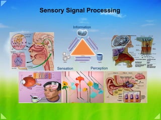
![Attributes of a Stimulus ,[object Object],[object Object],[object Object],[object Object]](data:image/gif;base64,R0lGODlhAQABAIAAAAAAAP///yH5BAEAAAAALAAAAAABAAEAAAIBRAA7)